

Preparation And Storage
Recommended Assay Procedures
BD® CompBeads can be used as surrogates to assess fluorescence spillover (compensation). When fluorochrome conjugated antibodies are bound to BD® CompBeads, they have spectral properties very similar to cells. However, for some fluorochromes there can be small differences in spectral emissions compared to cells, resulting in spillover values that differ when compared to biological controls. It is strongly recommended that when using a reagent for the first time, users compare the spillover on cells and BD® CompBeads to ensure that BD® CompBeads are appropriate for your specific cellular application.
Product Notices
- Please refer to www.bdbiosciences.com/us/s/resources for technical protocols.
- Caution: Sodium azide yields highly toxic hydrazoic acid under acidic conditions. Dilute azide compounds in running water before discarding to avoid accumulation of potentially explosive deposits in plumbing.
- This reagent has been pre-diluted for use at the recommended Volume per Test. We typically use 1 × 10^6 cells in a 100-µl experimental sample (a test).
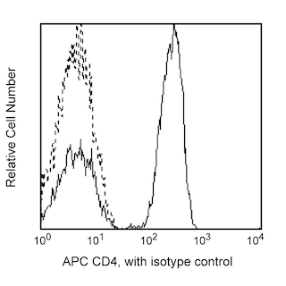
- An isotype control should be used at the same concentration as the antibody of interest.
- CF™ is a trademark of Biotium, Inc.
- Please refer to http://regdocs.bd.com to access safety data sheets (SDS).
- Human donor specific background has been observed in relation to the presence of anti-polyethylene glycol (PEG) antibodies, developed as a result of certain vaccines containing PEG, including some COVID-19 vaccines. We recommend use of BD Horizon Brilliant™ Stain Buffer in your experiments to help mitigate potential background. For more information visit https://www.bdbiosciences.com/en-us/support/product-notices.
- For fluorochrome spectra and suitable instrument settings, please refer to our Multicolor Flow Cytometry web page at www.bdbiosciences.com/colors.
Companion Products


The 2A3 monoclonal antibody specifically binds to human CD25, the low-affinity alpha subunit of the Interleukin-2 Receptor (IL- 2Rα). CD25 associates with CD122 (IL-2Rβ chain) and CD132 (common γ chain or γc) to form the high-affinity signal-transducing IL-2R complex. CD25 is expressed by subsets of thymocytes and peripheral blood lymphocytes including CD4+CD25+ regulatory T cells and memory T cells. CD25 antigen density increases on activated T cells including phytohemagglutinin (PHA)-, concanavalin A (Con A)-, and CD3-activated T lymphocytes. High levels of CD25 can be expressed by T lymphocytes from mixed lymphocyte cultures and by human T-lymphocyte leukemia virus (HTLV)-infected T-lymphocyte leukemia lines, for example, HUT-102. CD25 can also be expressed by activated B cells and macrophages. Recombinant IL-2 blocks the binding of the 2A3 antibody to PHA-activated T lymphocytes.
The BD Horizon RealYellow™ 586 (RY586) Dye is part of the BD family of yellow-green dyes. It is a small organic fluorochrome with an excitation maximum (Ex Max) at 565-nm and an emission maximum (Em Max) at 586-nm. Driven by BD innovation, RY586 can be used on both spectral and conventional cytometers and is designed to be excited by the Yellow-Green laser (561-nm) with minimal excitation by the 488-nm Blue laser. For conventional instruments equipped with a Yellow-Green laser (561-nm), RY586 can be used as an alternative to PE and we recommend using an optical filter centered near 586-nm (eg, a 586/15-nm bandpass filter). For spectral instruments equipped with a Yellow-Green laser (561-nm), it can be used in conjunction with PE. Compared to PE, RY586 is similar in brightness, minimal spillover into Blue detectors, and increased spillover into the 610/20-nm (PE-CF594) detector. Please ensure that your instrument configuration (lasers and optical filters) is appropriate for this dye.

Development References (12)
-
Dower SK, Hefeneider SH, Alpert AR, Urdal DL. Quantitative measurement of human interleukin 2 receptor levels with intact and detergent-solubilized human T-cells. Mol Immunol. 1985; 22(8):937-947. (Clone-specific). View Reference
-
Greene WC, Leonard WJ. The human interleukin-2 receptor. Annu Rev Immunol. 1986; 4:69-95. (Clone-specific). View Reference
-
Jackson AL, Matsumoto H, Janszen M, Maino V, Blidy A, Shye S. Restricted expression of p55 interleukin 2 receptor (CD25) on normal T cells. Clin Immunol Immunopathol. 1990; 54(1):126-133. (Clone-specific: Flow cytometry). View Reference
-
Leonard WJ, Depper JM, Uchiyama T, Smith KA, Waldmann TA, Greene WC. A monoclonal antibody that appears to recognize the receptor for human T-cell growth factor; partial characterization of the receptor. Nature. 1982; 300(5889):267-269. (Biology). View Reference
-
Ng WF, Duggan PJ, Ponchel F, et al. Human CD4(+)CD25(+) cells: a naturally occurring population of regulatory T cells. Blood. 2001; 98(9):2736-2744. (Biology). View Reference
-
Rambaldi A, Young DC, Herrmann F, Cannistra SA, Griffin JD. Interferon-gamma induces expression of the interleukin 2 receptor gene in human monocytes. Eur J Immunol. 1987; 17(1):153-156. (Clone-specific). View Reference
-
Robb RJ, Greene WC, Rusk CM. Low and high affinity cellular receptors for interleukin 2. Implications for the level of Tac antigen. J Exp Med. 1984; 160(4):1126-1146. (Biology). View Reference
-
Schwarting R, Stein H. Cluster report: CD25. In: Knapp W. W. Knapp .. et al., ed. Leucocyte typing IV : white cell differentiation antigens. Oxford New York: Oxford University Press; 1989:399-403.
-
Sereti I, Martinez-Wilson H, Metcalf JA, et al. Long-term effects of intermittent interleukin 2 therapy in patients with HIV infection: characterization of a novel subset of CD4(+)/CD25(+) T cells. Blood. 2002; 100(6):2159-2167. (Clone-specific: Flow cytometry). View Reference
-
Siegel JP, Sharon M, Smith PL, Leonard WJ. The IL-2 receptor beta chain (p70): role in mediating signals for LAK, NK, and proliferative activities. Science. 1987; 238(4823):75-78. (Biology). View Reference
-
Teshigawara K, Wang HM, Kato K, Smith KA. Interleukin 2 high-affinity receptor expression requires two distinct binding proteins. J Exp Med. 1987; 165(1):223-238. (Biology). View Reference
-
Urdal DL, March CJ, Gillis S, Larsen A, Dower SK. Purification and chemical characterization of the receptor for interleukin 2 from activated human T lymphocytes and from a human T-cell lymphoma cell line. Proc Natl Acad Sci U S A. 1984; 81(20):6481-6485. (Immunogen: Blocking, Dot Blot, Immunoaffinity chromatography, Inhibition, Radioimmunoassay). View Reference
Please refer to Support Documents for Quality Certificates
Global - Refer to manufacturer's instructions for use and related User Manuals and Technical data sheets before using this products as described
Comparisons, where applicable, are made against older BD Technology, manual methods or are general performance claims. Comparisons are not made against non-BD technologies, unless otherwise noted.
For Research Use Only. Not for use in diagnostic or therapeutic procedures.