BD OptiBuild™ RY586 Mouse Anti-Human CD183 (CXCR3)
Clone 1C6/CXCR3 (also known as 1C6, LS177-1C6)
(RUO)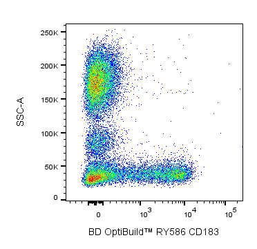
Preparation And Storage
Recommended Assay Procedures
BD® CompBeads can be used as surrogates to assess fluorescence spillover (compensation). When fluorochrome conjugated antibodies are bound to BD® CompBeads, they have spectral properties very similar to cells. However, for some fluorochromes there can be small differences in spectral emissions compared to cells, resulting in spillover values that differ when compared to biological controls. It is strongly recommended that when using a reagent for the first time, users compare the spillover on cells and BD® CompBeads to ensure that BD® CompBeads are appropriate for your specific cellular application.
Product Notices
- Researchers should determine the optimal concentration of this reagent for their individual applications.
- The production process underwent stringent testing and validation to assure that it generates a high-quality conjugate with consistent performance and specific binding activity. However, verification testing has not been performed on all conjugate lots.
- Please refer to www.bdbiosciences.com/us/s/resources for technical protocols.
- An isotype control should be used at the same concentration as the antibody of interest.
- Caution: Sodium azide yields highly toxic hydrazoic acid under acidic conditions. Dilute azide compounds in running water before discarding to avoid accumulation of potentially explosive deposits in plumbing.
- CF™ is a trademark of Biotium, Inc.
- Please refer to http://regdocs.bd.com to access safety data sheets (SDS).
- For fluorochrome spectra and suitable instrument settings, please refer to our Multicolor Flow Cytometry web page at www.bdbiosciences.com/colors.
- Since applications vary, each investigator should titrate the reagent to obtain optimal results.
- Human donor specific background has been observed in relation to the presence of anti-polyethylene glycol (PEG) antibodies, developed as a result of certain vaccines containing PEG, including some COVID-19 vaccines. We recommend use of BD Horizon Brilliant™ Stain Buffer in your experiments to help mitigate potential background. For more information visit https://www.bdbiosciences.com/en-us/support/product-notices.
Companion Products


The 1C6/CXCR3 monoclonal antibody specifically binds to human CD183, also known as the CXCR3 chemokine receptor. CD183 is a 40-41 kDa seven-transmembrane protein and member of the G protein-coupled receptor family. CD183 is expressed primarily on activated T cells that infiltrate inflammatory sites. It has also been detected on some circulating T cells, B cells, and NK cells. Reports show that some CXCR3-positive T cells also express CCR5 and are mostly CD45RO-positive cells. Three ligands for CXCR3 have been identified. They are CXCL9 (Mig/monokine induced by interferon-γ), CXCL10 (IP-10/interferon-γ inducible 10-kD protein), and CXCL11 (I-TAC/interferon-inducible T-cell alpha chemoattractant). These chemokines are produced by a variety of cells upon stimulation by IFN-γ and interact with CXCR3 to mediate T-cell chemotaxis. This reagent has been reported to be suitable for immunohistochemical staining of acetone-fixed, frozen sections and/or formalin-fixed, paraffin-embedded tissue sections with citrate pretreatment. Clone 1C6/CXCR3 also cross reacts with a subset of peripheral blood lymphocytes of baboon, and both rhesus and cynomolgus macaque monkeys. The distribution of lymphocytes is similar to that observed with CD183-positive peripheral blood lymphocytes from normal human donors. CXCR3 has been clustered as CD183 in the VIIth HLDA workshop.

Development References (5)
-
Loetscher M, Gerber B, Loetscher P, et al. Chemokine receptor specific for IP10 and mig: structure, function, and expression in activated T-lymphocytes. J Exp Med. 1996; 184(3):963-969. (Biology). View Reference
-
Marcher C, Moller BK, Lillevang ST, Kristensen T. CXCR4 and IL17R are downregulated on cord-blood CD34-positive cells during short-term culture. In: Mason D. David Mason .. et al., ed. Leucocyte typing VII : white cell differentiation antigens : proceedings of the Seventh International Workshop and Conference held in Harrogate, United Kingdom. Oxford: Oxford University Press; 2002:629-632.
-
Piali L, Weber C, LaRosa G, et al. The chemokine receptor CXCR3 mediates rapid and shear-resistant adhesion-induction of effector T lymphocytes by the chemokines IP10 and Mig. Eur J Immunol. 1998; 28(3):961-972. (Biology). View Reference
-
Qin S, Rottman JB, Myers P, et al. The chemokine receptors CXCR3 and CCR5 mark subsets of T cells associated with certain inflammatory reactions. J Clin Invest. 1998; 101(4):746-754. (Immunogen: Blocking, Flow cytometry, Immunohistochemistry, Inhibition). View Reference
-
Uguccioni M, Willimann K. Cytokine/Chemokine Receptors: Section report. In: Mason D. David Mason .. et al., ed. Leucocyte typing VII : white cell differentiation antigens : proceedings of the Seventh International Workshop and Conference held in Harrogate, United Kingdom. Oxford: Oxford University Press; 2002:237-243.
Please refer to Support Documents for Quality Certificates
Global - Refer to manufacturer's instructions for use and related User Manuals and Technical data sheets before using this products as described
Comparisons, where applicable, are made against older BD Technology, manual methods or are general performance claims. Comparisons are not made against non-BD technologies, unless otherwise noted.
For Research Use Only. Not for use in diagnostic or therapeutic procedures.