Skip to main content Skip to navigation
RY586 Mouse Anti-Human CD152
Multiparameter flow cytometric analysis of CD152 expression on PHA-activated Human peripheral blood lymphocytes. Human peripheral blood mononuclear cells stimulated with Phytohemagglutinin (PHA) for 3 days were preincubated with BD Pharmingen™ Human BD Fc Block™ (Cat. No. 564219/564220). These cells were then stained with BD Horizon™ R718 Mouse Anti-Human CD3 antibody (Cat. No. 566953/566954) and with either BD Horizon™ RY586 Mouse IgG2a, κ Isotype Control (Cat. No. 568131; Left Plot) or BD Horizon™ RY586 Mouse Anti-Human CD152 antibody (Cat. No. 568441/568442; Right Plot). BD Via-Probe™ Cell Viability 7-AAD Solution (Cat. No. 555815/555816) was added to cells right before analysis. The bivariate pseudocolor density plot showing the correlated expression of CD152 (or Ig Isotype control staining) versus CD3 was derived from gated events with the forward and side light-scatter characteristics of viable (7-AAD-negative) lymphocytes. Flow cytometry and data analysis were performed using a BD LSRFortessa™ X-20 Cell Analyzer System and FlowJo™ software.
RY586 Mouse Anti-Human CD152
Multiparameter flow cytometric analysis of CD152 expression on Con A-activated Human peripheral blood lymphocytes. Human peripheral blood mononuclear cells stimulated with Concanavalin A (Con A) for 3 days were preincubated with BD Pharmingen™ Human BD Fc Block™ (Cat. No. 564219/564220). These cells were then stained with BD Horizon™ R718 Mouse Anti-Human CD3 antibody (Cat. No. 566953/566954) and with either BD Horizon™ RY586 Mouse IgG2a, κ Isotype Control (Cat. No. 568131; Left Plot) or BD Horizon™ RY586 Mouse Anti-Human CD152 antibody (Cat. No. 568441/568442; Right Plot). BD Via-Probe™ Cell Viability 7-AAD Solution (Cat. No. 555815/555816) was added to cells right before analysis. The bivariate pseudocolor density plot showing the correlated expression of CD152 (or Ig Isotype control staining) versus CD3 was derived from gated events with the forward and side light-scatter characteristics of viable (7-AAD-negative) lymphocytes. Flow cytometry and data analysis were performed using a BD LSRFortessa™ X-20 Cell Analyzer System and FlowJo™ software.
Multiparameter flow cytometric analysis of CD152 expression on PHA-activated Human peripheral blood lymphocytes. Human peripheral blood mononuclear cells stimulated with Phytohemagglutinin (PHA) for 3 days were preincubated with BD Pharmingen™ Human BD Fc Block™ (Cat. No. 564219/564220). These cells were then stained with BD Horizon™ R718 Mouse Anti-Human CD3 antibody (Cat. No. 566953/566954) and with either BD Horizon™ RY586 Mouse IgG2a, κ Isotype Control (Cat. No. 568131; Left Plot) or BD Horizon™ RY586 Mouse Anti-Human CD152 antibody (Cat. No. 568441/568442; Right Plot). BD Via-Probe™ Cell Viability 7-AAD Solution (Cat. No. 555815/555816) was added to cells right before analysis. The bivariate pseudocolor density plot showing the correlated expression of CD152 (or Ig Isotype control staining) versus CD3 was derived from gated events with the forward and side light-scatter characteristics of viable (7-AAD-negative) lymphocytes. Flow cytometry and data analysis were performed using a BD LSRFortessa™ X-20 Cell Analyzer System and FlowJo™ software.
Multiparameter flow cytometric analysis of CD152 expression on Con A-activated Human peripheral blood lymphocytes. Human peripheral blood mononuclear cells stimulated with Concanavalin A (Con A) for 3 days were preincubated with BD Pharmingen™ Human BD Fc Block™ (Cat. No. 564219/564220). These cells were then stained with BD Horizon™ R718 Mouse Anti-Human CD3 antibody (Cat. No. 566953/566954) and with either BD Horizon™ RY586 Mouse IgG2a, κ Isotype Control (Cat. No. 568131; Left Plot) or BD Horizon™ RY586 Mouse Anti-Human CD152 antibody (Cat. No. 568441/568442; Right Plot). BD Via-Probe™ Cell Viability 7-AAD Solution (Cat. No. 555815/555816) was added to cells right before analysis. The bivariate pseudocolor density plot showing the correlated expression of CD152 (or Ig Isotype control staining) versus CD3 was derived from gated events with the forward and side light-scatter characteristics of viable (7-AAD-negative) lymphocytes. Flow cytometry and data analysis were performed using a BD LSRFortessa™ X-20 Cell Analyzer System and FlowJo™ software.
Product Details
Down Arrow Up Arrow


BD Horizon™
CTLA-4; AILIM; Cytotoxic T-lymphocyte protein 4
Human (QC Testing), Rhesus,Cynomolgus,Baboon (Tested in Development)
Mouse BALB/c IgG2a, κ
Human CTLA4 Recombinant Protein
Flow cytometry (Routinely Tested), Intracellular staining (flow cytometry) (Tested During Development)
5 µl
IX 34
1493
Aqueous buffered solution containing ≤0.09% sodium azide.
RUO


Preparation And Storage

Store undiluted at 4°C and protected from prolonged exposure to light. Do not freeze. The monoclonal antibody was purified from tissue culture supernatant or ascites by affinity chromatography. The antibody was conjugated to the dye under optimum conditions and unreacted dye was removed.

Recommended Assay Procedures

BD® CompBeads can be used as surrogates to assess fluorescence spillover (compensation). When fluorochrome conjugated antibodies are bound to BD® CompBeads, they have spectral properties very similar to cells. However, for some fluorochromes there can be small differences in spectral emissions compared to cells, resulting in spillover values that differ when compared to biological controls. It is strongly recommended that when using a reagent for the first time, users compare the spillover on cells and BD® CompBeads to ensure that BD® CompBeads are appropriate for your specific cellular application.

Product Notices

  1. Please refer to www.bdbiosciences.com/us/s/resources for technical protocols.
  2. This reagent has been pre-diluted for use at the recommended Volume per Test. We typically use 1 × 10^6 cells in a 100-µl experimental sample (a test).
  3. An isotype control should be used at the same concentration as the antibody of interest.
  4. Caution: Sodium azide yields highly toxic hydrazoic acid under acidic conditions. Dilute azide compounds in running water before discarding to avoid accumulation of potentially explosive deposits in plumbing.
  5. For fluorochrome spectra and suitable instrument settings, please refer to our Multicolor Flow Cytometry web page at www.bdbiosciences.com/colors.
  6. Human donor specific background has been observed in relation to the presence of anti-polyethylene glycol (PEG) antibodies, developed as a result of certain vaccines containing PEG, including some COVID-19 vaccines. We recommend use of BD Horizon Brilliant™ Stain Buffer in your experiments to help mitigate potential background. For more information visit https://www.bdbiosciences.com/en-us/support/product-notices.
  7. Please refer to http://regdocs.bd.com to access safety data sheets (SDS).
  8. CF™ is a trademark of Biotium, Inc.
  9. For U.S. patents that may apply, see bd.com/patents.
568441 Rev. 2
Antibody Details
Down Arrow Up Arrow
BNI3

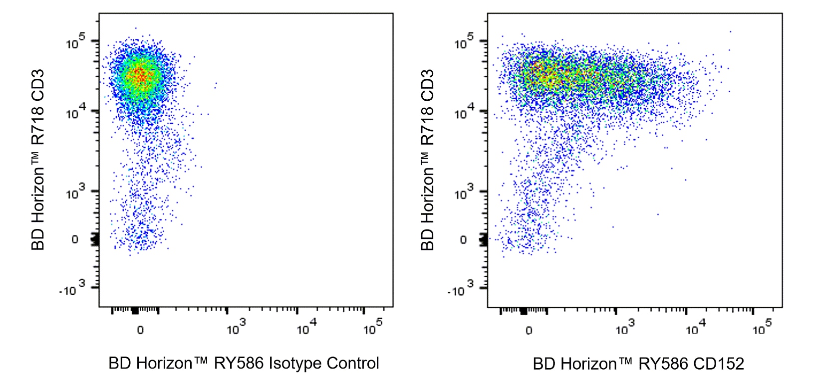
The BNI3 monoclonal antibody specifically binds to the human cytolytic T lymphocyte-associated antigen (CTLA-4), also known as CD152. CTLA-4 is transiently expressed on activated CD28+ T cells and binds to CD80 and CD86 present on antigen presenting cells (APC) with high avidity. This interaction appears to deliver a negative regulatory signal to the T cell. Recent reports indicate that CTLA-4 is also expressed on B cells when cultured with activated T cells, suggesting a role for CTLA-4 in the regulation of B-cell response. Immobilized BNI3 antibody enhances T-cell proliferation induced by antibody-mediated crosslinking of CD3 and CD28. Recent studies have shown that CD152 can be expressed by regulatory T (Treg) cells. After cellular fixation and permeabilization, the BNI3 antibody can stain intracellular CD152 expressed in T cells including Treg cells. Clone BNI3 was studied in the VI Leukocyte Typing Workshop.

568441 Rev. 2
Format Details
Down Arrow Up Arrow
RY586
The BD Horizon RealYellow™ 586 (RY586) Dye is part of the BD family of yellow-green dyes. It is a small organic fluorochrome with an excitation maximum (Ex Max) at 565-nm and an emission maximum (Em Max) at 586-nm. Driven by BD innovation, RY586 can be used on both spectral and conventional cytometers and is designed to be excited by the Yellow-Green laser (561-nm) with minimal excitation by the 488-nm Blue laser. For conventional instruments equipped with a Yellow-Green laser (561-nm), RY586 can be used as an alternative to PE and we recommend using an optical filter centered near 586-nm (eg, a 586/15-nm bandpass filter). For spectral instruments equipped with a Yellow-Green laser (561-nm), it can be used in conjunction with PE. Compared to PE, RY586 is similar in brightness, minimal spillover into Blue detectors, and increased spillover into the 610/20-nm (PE-CF594) detector. Please ensure that your instrument configuration (lasers and optical filters) is appropriate for this dye.
altImg
RY586
Yellow-Green 561 nm
564 nm
586 nm
568441 Rev.2
Citations & References
Down Arrow Up Arrow

Development References (10)

  1. Cabezon R, Sintes J, Llinas L, Benitez-Ribas D. Analysis of HLDA9 mAbs on plasmacytoid dendritic cell. Immunol Lett. 2011; 134(2):167-173. (Clone-specific: Flow cytometry). View Reference
  2. Castan J, Klauenberg U, Kalmar P, Fleischer B, Broker BM. Expression of CTLA-4 (CD152) on human medullary CD4+ thymocytes. Med Microbiol Immunol (Berl). 1998; 187(1):49-52. (Immunogen: Fluorescence microscopy, Immunocytochemistry, Immunofluorescence, Immunohistochemistry). View Reference
  3. Castan J, Tenner-Racz K, Racz P, Fleischer B, Broker BM. Accumulation of CTLA-4 expressing T lymphocytes in the germinal centres of human lymphoid tissues. Immunology. 1997; 90(2):265-271. (Immunogen: ELISA, Fluorescence microscopy, Immunofluorescence, Immunohistochemistry). View Reference
  4. Healy ZR, Murdoch DM. OMIP-036: Co-inhibitory receptor (immune checkpoint) expression analysis in human T cell subsets.. Cytometry A. 2016; 89(10):889-892. (Clone-specific: Intracellular Staining/Flow Cytometry). View Reference
  5. Kuiper HM, Brouwer M, Linsley PS, van Lier RA. Activated T cells can induce high levels of CTLA-4 expression on B cells. J Immunol. 1995; 155(4):1776-1783. (Biology). View Reference
  6. Lindsten T, Lee KP, Harris ES, et al. Characterization of CTLA-4 structure and expression on human T cells. J Immunol. 1993; 151(7):3489-3499. (Biology). View Reference
  7. Morton PA, Fu XT, Stewart JA, et al. Differential effects of CTLA-4 substitutions on the binding of human CD80 (B7-1) and CD86 (B7-2). J Immunol. 1996; 156(3):1047-1054. (Biology). View Reference
  8. Rabe H, Lundell AC, Andersson K, Adlerberth I, Wold AE, Rudin A. Higher proportions of circulating FOXP3+ and CTLA-4+ regulatory T cells are associated with lower fractions of memory CD4+ T cells in infants.. J Leukoc Biol. 2011; 90(6):1133-40. (Clone-specific: Intracellular Staining/Flow Cytometry). View Reference
  9. Santegoets SJ, Dijkgraaf EM, Battaglia A, et al. Monitoring regulatory T cells in clinical samples: consensus on an essential marker set and gating strategy for regulatory T cell analysis by flow cytometry.. Cancer Immunol Immunother. 2015; 64(10):1271-86. (Clone-specific: Intracellular Staining/Flow Cytometry). View Reference
  10. Wang H, Shih CC, Waters JB, et al. CD152 (CTLA4) Workshop: Expression and function of CD152 on human T cells: A study using a mouse anti-human CD152 monoclonal antibody BNI3.1. In: Kishimoto T. Tadamitsu Kishimoto .. et al., ed. Leucocyte typing VI : white cell differentiation antigens : proceedings of the sixth international workshop and conference held in Kobe, Japan, 10-14 November 1996. New York: Garland Pub.; 1997:97-98.
View All (10) View Less
568441 Rev. 2

Please refer to Support Documents for Quality Certificates


Global - Refer to manufacturer's instructions for use and related User Manuals and Technical data sheets before using this products as described


Comparisons, where applicable, are made against older BD Technology, manual methods or are general performance claims.  Comparisons are not made against non-BD technologies, unless otherwise noted.

For Research Use Only. Not for use in diagnostic or therapeutic procedures.