Preparation And Storage
Recommended Assay Procedures
BD® CompBeads can be used as surrogates to assess fluorescence spillover (compensation). When fluorochrome conjugated antibodies are bound to BD® CompBeads, they have spectral properties very similar to cells. However, for some fluorochromes there can be small differences in spectral emissions compared to cells, resulting in spillover values that differ when compared to biological controls. It is strongly recommended that when using a reagent for the first time, users compare the spillover on cells and BD® CompBeads to ensure that BD® CompBeads are appropriate for your specific cellular application.
Product Notices
- Please refer to www.bdbiosciences.com/us/s/resources for technical protocols.
- Caution: Sodium azide yields highly toxic hydrazoic acid under acidic conditions. Dilute azide compounds in running water before discarding to avoid accumulation of potentially explosive deposits in plumbing.
- This reagent has been pre-diluted for use at the recommended Volume per Test. We typically use 1 × 10^6 cells in a 100-µl experimental sample (a test).
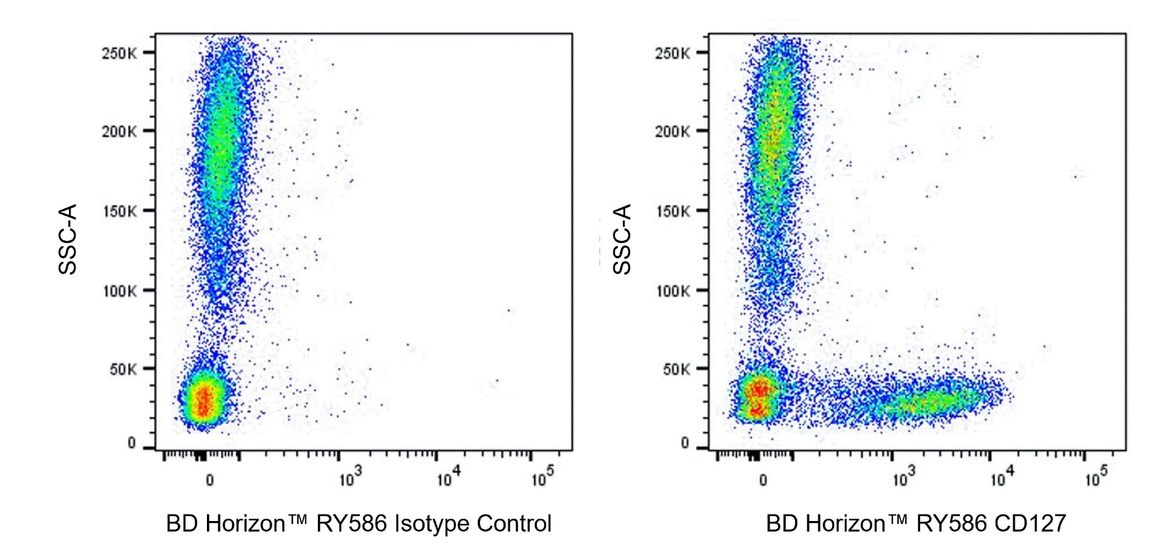
- An isotype control should be used at the same concentration as the antibody of interest.
- CF™ is a trademark of Biotium, Inc.
- Human donor specific background has been observed in relation to the presence of anti-polyethylene glycol (PEG) antibodies, developed as a result of certain vaccines containing PEG, including some COVID-19 vaccines. We recommend use of BD Horizon Brilliant™ Stain Buffer in your experiments to help mitigate potential background. For more information visit https://www.bdbiosciences.com/en-us/support/product-notices.
- Please refer to http://regdocs.bd.com to access safety data sheets (SDS).
- For fluorochrome spectra and suitable instrument settings, please refer to our Multicolor Flow Cytometry web page at www.bdbiosciences.com/colors.
Companion Products


The hIL-7R-M21 monoclonal antibody specifically binds to the 60-90 kDa glycoprotein, CD127. CD127 is also known as the IL-7 receptor alpha (IL-7Rα) subunit. The IL-7 receptor complex is a heterodimer composed of CD127 and the common gamma chain (γc, CD132), shared by other cytokine receptors (IL-2R, IL-4R, IL-9R, IL-15R, and IL-21R). CD127 is expressed on thymocytes, T- and B-cell progenitors, mature T cells, and some lymphoid and myeloid cells. In vitro experiments show the expression of CD127 is down-regulated following T cell activation. Studies indicate that the IL-7 Receptor plays an important role in the proliferation and differentiation of mature T cells. Recently, it has been shown that low surface expression of CD127, in combination with intermediate to high surface expression of CD25, the α chain of the IL-2 receptor complex, can distinguish between human regulatory and conventional CD4+ T cells in human adult and cord blood, lymph nodes and thymus.
The BD Horizon RealYellow™ 586 (RY586) Dye is part of the BD family of yellow-green dyes. It is a small organic fluorochrome with an excitation maximum (Ex Max) at 565-nm and an emission maximum (Em Max) at 586-nm. Driven by BD innovation, RY586 can be used on both spectral and conventional cytometers and is designed to be excited by the Yellow-Green laser (561-nm) with minimal excitation by the 488-nm Blue laser. For conventional instruments equipped with a Yellow-Green laser (561-nm), RY586 can be used as an alternative to PE and we recommend using an optical filter centered near 586-nm (eg, a 586/15-nm bandpass filter). For spectral instruments equipped with a Yellow-Green laser (561-nm), it can be used in conjunction with PE. Compared to PE, RY586 is similar in brightness, minimal spillover into Blue detectors, and increased spillover into the 610/20-nm (PE-CF594) detector. Please ensure that your instrument configuration (lasers and optical filters) is appropriate for this dye.

Development References (8)
-
Appasamy PM. Biological and clinical implications of interleukin-7 and lymphopoiesis. Cytokines Cell Mol Ther. 1999; 5(1):25-39. (Biology: Flow cytometry). View Reference
-
Armitage RJ, Ziegler SF, Friend DJ, Park LS, Fanslow WC. Identification of a novel low-affinity receptor for human interleukin-7. Blood. 1992; 79(7):1738-1745. (Immunogen: Flow cytometry). View Reference
-
Benjamin D, Sharma V, Knobloch TJ, Armitage RJ, Dayton MA, Goodwin RG. B cell IL-7. Human B cell lines constitutively secrete IL-7 and express IL-7 receptors. J Immunol. 1994; 152(10):4749-4757. (Clone-specific: Flow cytometry). View Reference
-
Goodwin RG, Friend D, Ziegler SF et al. Cloning of the human and murine interleukin-7 receptors: demonstration of a soluble form and homology to a new receptor superfamily. Cell. 1990; 60(6):941-951. (Biology). View Reference
-
Hofmeister R, Khaled AR, Benbernou N, Rajnavolgyi E, Muegge K, Durum SK. Interleukin-7: physiological roles and mechanisms of action. Cytokine Growth Factor Rev. 1999; 10(1):41-60. (Biology). View Reference
-
Liu W, Putnam AL, Xu-Yu Z, et al. CD127 expression inversely correlates with FoxP3 and suppressive function of human CD4+ T reg cells. J Exp Med. 2006; 203(7):1701-1711. (Clone-specific: Flow cytometry, Fluorescence activated cell sorting). View Reference
-
Sasidharan Nair V, Toor SM, Taouk G, et al. Pembrolizumab Interferes with the Differentiation of Human FOXP3(+)-Induced T Regulatory Cells, but Not with FOXP3 Stability, through Activation of mTOR. J Immunol. 2020; 204(1):199-211. (Clone-specific: Flow cytometry, Fluorescence activated cell sorting). View Reference
-
Seddiki N, Santner-Nanan B, Martinson J et al. Expression of interleukin (IL)-2 and IL-7 receptors discriminates between human regulatory and activated T cells. J Exp Med. 2006; 203(7):1693-1700. (Biology). View Reference
Please refer to Support Documents for Quality Certificates
Global - Refer to manufacturer's instructions for use and related User Manuals and Technical data sheets before using this products as described
Comparisons, where applicable, are made against older BD Technology, manual methods or are general performance claims. Comparisons are not made against non-BD technologies, unless otherwise noted.
For Research Use Only. Not for use in diagnostic or therapeutic procedures.