-
Reagents
- Flow Cytometry Reagents
-
Western Blotting and Molecular Reagents
- Immunoassay Reagents
-
Single-Cell Multiomics Reagents
- BD® AbSeq Assay
- BD Rhapsody™ Accessory Kits
- BD® Single-Cell Multiplexing Kit
- BD Rhapsody™ Targeted mRNA Kits
- BD Rhapsody™ Whole Transcriptome Analysis (WTA) Amplification Kit
- BD Rhapsody™ TCR/BCR Profiling Assays for Human and Mouse
- BD® OMICS-Guard Sample Preservation Buffer
- BD Rhapsody™ ATAC-Seq Assays
-
Functional Assays
-
Microscopy and Imaging Reagents
-
Cell Preparation and Separation Reagents
-
Training
- Flow Cytometry Basic Training
-
Product-Based Training
- BD FACSDiscover™ S8 Cell Sorter Product Training
- Accuri C6 Plus Product-Based Training
- FACSAria Product Based Training
- FACSCanto Product-Based Training
- FACSLyric Product-Based Training
- FACSMelody Product-Based Training
- FACSymphony Product-Based Training
- HTS Product-Based Training
- LSRFortessa Product-Based Training
- Advanced Training
-
- BD® AbSeq Assay
- BD Rhapsody™ Accessory Kits
- BD® Single-Cell Multiplexing Kit
- BD Rhapsody™ Targeted mRNA Kits
- BD Rhapsody™ Whole Transcriptome Analysis (WTA) Amplification Kit
- BD Rhapsody™ TCR/BCR Profiling Assays for Human and Mouse
- BD® OMICS-Guard Sample Preservation Buffer
- BD Rhapsody™ ATAC-Seq Assays
-
- BD FACSDiscover™ S8 Cell Sorter Product Training
- Accuri C6 Plus Product-Based Training
- FACSAria Product Based Training
- FACSCanto Product-Based Training
- FACSLyric Product-Based Training
- FACSMelody Product-Based Training
- FACSymphony Product-Based Training
- HTS Product-Based Training
- LSRFortessa Product-Based Training
- United States (English)
-
Change country/language
Old Browser
This page has been recently translated and is available in French now.
Looks like you're visiting us from {countryName}.
Would you like to stay on the current country site or be switched to your country?

Standard curve mouse MCP-1


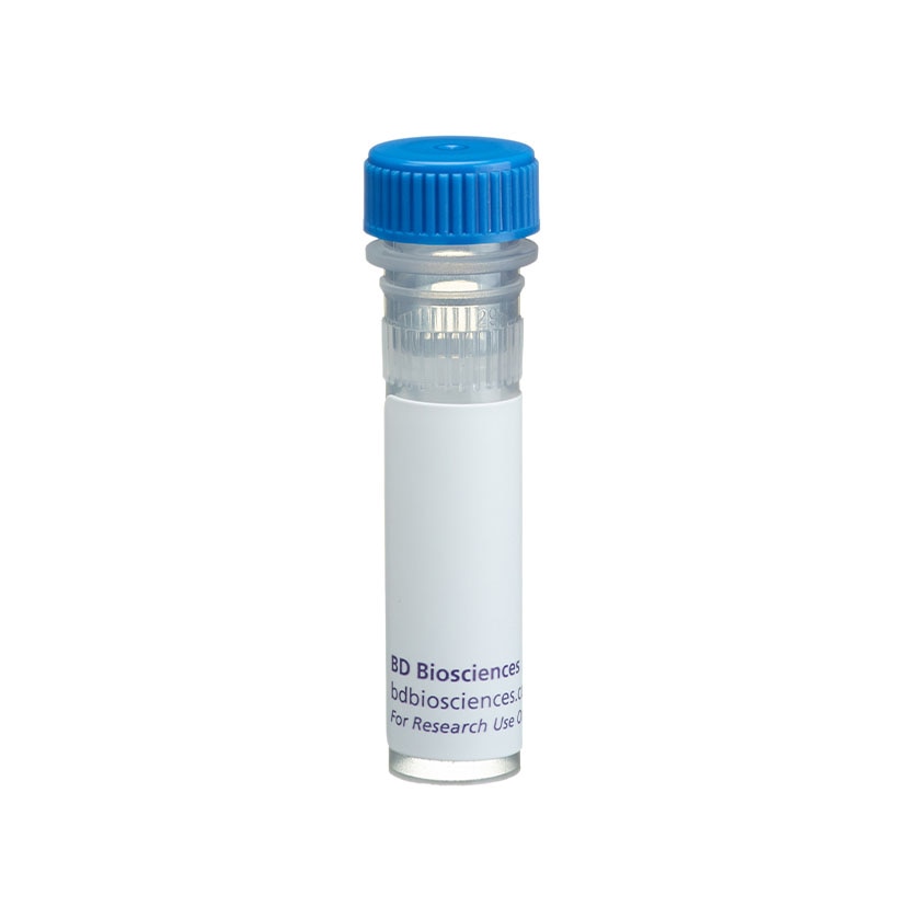
BD Pharmingen™ Purified Hamster Anti-Mouse MCP-1

Regulatory Status Legend
Any use of products other than the permitted use without the express written authorization of Becton, Dickinson and Company is strictly prohibited.
Preparation And Storage
Recommended Assay Procedures
ELISA: The purified 2H5 antibody (Cat. No. 551217) is useful as a capture antibody for a sandwich ELISA for measuring mouse MCP-1 protein levels. Purified 2H5 antibody can be paired with the biotinylated 4E2/MCP antibody (Cat. No. 554444) as the detecting antibody, with recombinant mouse MCP-1 (Cat. No. 554590) as the standard. Purified 2H5 antibody should be titrated 6 - 10 µg/ml to determine optimal concentration for ELISA capture. To obtain linear standard curves, doubling dilutions of mouse MCP-1 ranging from ~4,000 to 30 pg/ml are recommended for inclusion in each ELISA plate. For maximal sensitivity, an overnight incubation (4°C) of samples/standards with the coated capture antibody is recommended. For specific methodology, please visit the protocols section of the Immune Function Handbook, which is also posted on our web site, www.bdbiosciences.com.
Note 1: This reagent is recommended primarily for assay of cytokine from experimental cell culture systems. For detection of MCP-1 in mouse serum or plasma samples, the BD OptEIA™ Mouse MCP-1 ELISA Set is recommended.
Note 2: This ELISA pair shows no cross-reactivity with any of the cytokines tested (e.g., mouse IL-1β, IL-2, IL-3, IL-4, IL-5, IL-6, IL-7, IL-9, IL-10, IL-12 p70, IL-15, GM-CSF, IFN-γ, TCA-3, TNF; human IL-1α, IL-1β, IL-2, IL-3, IL-4, IL-5, IL-6, IL-7, IL-8, IL-9, IL-10, IL-11, IL-12 p70, IL-12 p40, IL-13, IL-15, G-CSF, GM-CSF, IFN-γ, lymphotactin, MCP-1, MCP-2, MIP-1α, MIP-1β, NT-3, PDGF-AA, sCD23 , SCF, TNF, TNF-β, VEGF; rat IL-2, IL-4, IL-6, IL-10, GM-CSF, IFN-γ, TNF).
Neutralization: The NA/LE™ 2H5 antibody (Cat. No. 554440) is useful for neutralization of mouse MCP-1 bioactivity. A suitable NA/LE™ hamster IgG isotype-matched control is the G235-2356 antibody (Cat. No. 554709).
Immunofluorescent Staining and Flow Cytometric Analysis: The 2H5 antibody is useful for immunofluorescent staining and flow cytometric analysis to identify and enumerate MCP-1 producing cells within mixed cell populations. PE conjugated 2H5 antibody (Cat. No. 554443) is especially suitable for these experiments. The use of a specificity control, such as recombinant mouse MCP-1 (Cat No. 554590) or unlabeled 2H5 antibody (Cat. No. 554441) is recommended. For specific methodology, please visit the protocols section or the Immune Function Handbook, which is also posted on our web site, www.bdbiosciences.com.
Product Notices
- Since applications vary, each investigator should titrate the reagent to obtain optimal results.
- Please refer to www.bdbiosciences.com/us/s/resources for technical protocols.
- Although hamster immunoglobulin isotypes have not been well defined, BD Biosciences Pharmingen has grouped Armenian and Syrian hamster IgG monoclonal antibodies according to their reactivity with a panel of mouse anti-hamster IgG mAbs. A table of the hamster IgG groups, Reactivity of Mouse Anti-Hamster Ig mAbs, may be viewed at http://www.bdbiosciences.com/documents/hamster_chart_11x17.pdf.
- Caution: Sodium azide yields highly toxic hydrazoic acid under acidic conditions. Dilute azide compounds in running water before discarding to avoid accumulation of potentially explosive deposits in plumbing.
The 2H5 antibody reacts with mouse and rat monocyte chemoattractant protein (MCP-1), formerly termed JE. This antibody also recognizes human MCP-1, but shows no reactivity with the closely related mouse β chemokines, TCA3 and MIP-1β. The immunogen used to generate the 2H5 hybridoma was heparin-purified CHO-expressed mouse MCP-1. This is a neutralizing antibody.
Development References (3)
-
Prussin C, Metcalfe DD. Detection of intracytoplasmic cytokine using flow cytometry and directly conjugated anti-cytokine antibodies. J Immunol Methods. 1995; 188(1):117-128. (Methodology: IC/FCM Block). View Reference
-
Sakanashi Y, Takeya M, Yoshimura T, Feng L, Morioka T, Takahashi K. Kinetics of macrophage subpopulations and expression of monocyte chemoattractant protein-1 (MCP-1) in bleomycin-induced lung injury of rats studied by a novel monoclonal antibody against rat MCP-1. J Leukoc Biol. 1994; 56(6):741-750. (Biology). View Reference
-
Yoshimura T, Takeya M, Takahashi K. Molecular cloning of rat monocyte chemoattractant protein-1 (MCP-1) and its expression in rat spleen cells and tumor cell lines. Biochem Biophys Res Commun. 1991; 174(2):504-509. (Clone-specific: Neutralization). View Reference
Please refer to Support Documents for Quality Certificates
Global - Refer to manufacturer's instructions for use and related User Manuals and Technical data sheets before using this products as described
Comparisons, where applicable, are made against older BD Technology, manual methods or are general performance claims. Comparisons are not made against non-BD technologies, unless otherwise noted.
For Research Use Only. Not for use in diagnostic or therapeutic procedures.
Report a Site Issue
This form is intended to help us improve our website experience. For other support, please visit our Contact Us page.