Dear Customer, If you experience any text readability or display issues on our website, please update your web browser to the latest version
.
See More
Dear Customer, If you experience any text readability or display issues on our website, please update your web browser to the latest version
.
See MoreDear Customer, If you experience any text readability or display issues on our website, please update your web browser to the latest version
.
This page has been recently translated and is available in French now.
Looks like you're visiting us from {countryName}.
Would you like to stay on the current location site or be switched to your location?
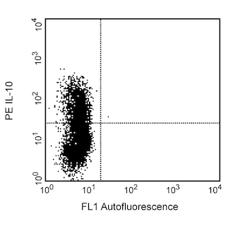
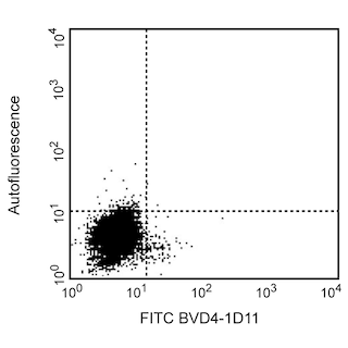
Expression of IL-4 by stimulated CD4+ and CD4-BALB/c spleen cells. Splenocytes from 6 month old BALB/c mice were cultured for 72 hours in medium containing Staphylococcus aureus enterotoxin B (2 µg/mL final concentration; Sigma, St. Louis, MO), recombinant mouse IL-2 (10 U/mL final concentration; Cat. No. 550069) and recombinant mouse IL-4 (2 ng/mL final concentration; Cat. No. 550067). The cells were harvested and restimulated for 5 hours with anti-CD3 (2 µg/mL final concentration; 145-2C11, Cat. No. 553057) and anti-CD28 (2 µg/mL final concentration; clone 37.51, Cat. No. 553294) antibodies in the presence of GolgiStop™ (2 µM final concentration; (Cat. No. 554724). The splenocytes were harvested, stained with 0.06 µg of FITC-conjugated rat anti-mouse CD4 (FITC-RM4-5, Cat. No. 553047) fixed, permeabilized, and subsequently stained with 0.25 µg of PE-conjugated rat anti-mouse IL-4 antibody (PE-BVD4-1D11, Cat. No. 554389) by using the BD Pharmingen staining protocol (see left panel). To demonstrate specificity of staining, the binding of PE-BVD4-1D11 was blocked by the preincubation of the conjugated antibody with recombinant mouse IL-4 (0.25 µg, Cat. No. 550067; see middle panel), and by preincubation of the fixed/permeabilized cells with unlabelled BVD4-1D11 antibody (5 µg, Cat. No. 554386; see right panel). The quadrant markers for the bivariate dot plots were set based on the autofluorescence control, and verified with the recombinant cytokine blocking and unlabelled antibody blocking specificity controls.
Any use of products other than the permitted use without the express written authorization of Becton, Dickinson and Company is strictly prohibited.
IL-4 is a pleiotropic cytokine that can regulate immunological responses including cell proliferation, survival and gene expression. Expressed by mast cells, basophils, eosinophils and T-cells, IL-4 regulates the differentiation of naive CD4+ T cells into helper Th2 cells. IL-4 can also be involved with the regulation of immunoglobulin class switching to the IgG1 and IgE isotypes in addition to being involved with the development of allergic inflammation and asthma.

Please refer to Support Documents for Quality Certificates
Global - Refer to manufacturer's instructions for use and related User Manuals and Technical data sheets before using this products as described
Comparisons, where applicable, are made against older BD Technology, manual methods or are general performance claims. Comparisons are not made against non-BD technologies, unless otherwise noted.
For Research Use Only. Not for use in diagnostic or therapeutic procedures.