Preparation And Storage
Recommended Assay Procedures
BD® CompBeads can be used as surrogates to assess fluorescence spillover (compensation). When fluorochrome conjugated antibodies are bound to BD® CompBeads, they have spectral properties very similar to cells. However, for some fluorochromes there can be small differences in spectral emissions compared to cells, resulting in spillover values that differ when compared to biological controls. It is strongly recommended that when using a reagent for the first time, users compare the spillover on cell and BD® CompBeads to ensure that BD® CompBeads are appropriate for your specific cellular application.
Product Notices
- Please refer to www.bdbiosciences.com/us/s/resources for technical protocols.
- Caution: Sodium azide yields highly toxic hydrazoic acid under acidic conditions. Dilute azide compounds in running water before discarding to avoid accumulation of potentially explosive deposits in plumbing.
- For fluorochrome spectra and suitable instrument settings, please refer to our Multicolor Flow Cytometry web page at www.bdbiosciences.com/colors.
- Please refer to http://regdocs.bd.com to access safety data sheets (SDS).
- Since applications vary, each investigator should titrate the reagent to obtain optimal results.
- An isotype control should be used at the same concentration as the antibody of interest.
Companion Products


.png?imwidth=320)


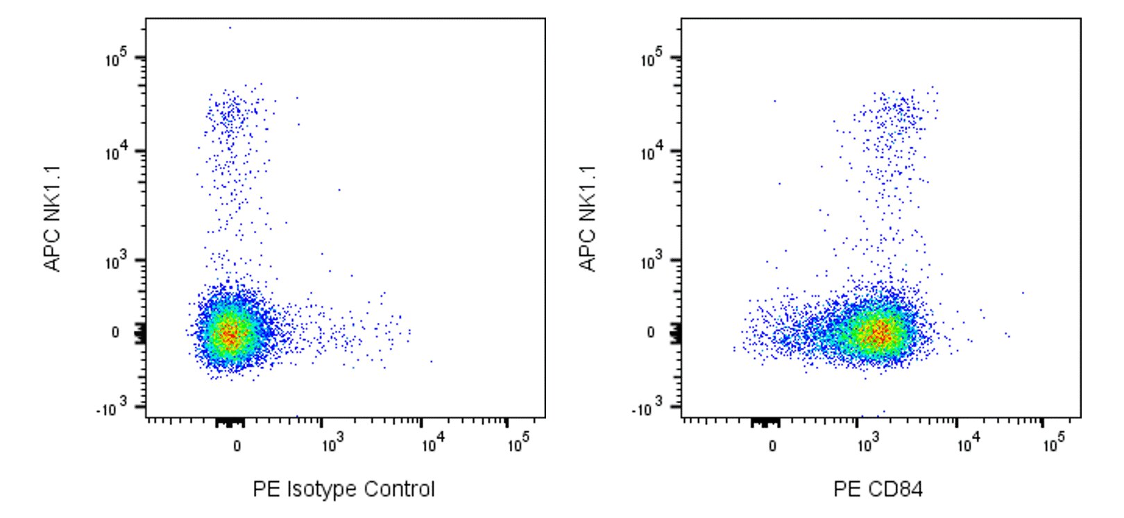
The mCD84.7.rMAb is a recombinant monoclonal antibody that was derived from mCD84.7 hybridoma cells. This antibody specifically recognizes CD84 which is also known as Signaling lymphocytic activation molecule 5 (SLAM family member 5 or Slamf5). CD84 is a single-pass type I transmembrane glycoprotein that is encoded by Cd84 (CD84 antigen) which belongs to the SLAM family within the CD2-subset of the immunoglobulin superfamily (IgSF) of cell surface receptors. CD84 is comprised of an extracellular region with one N-terminal Ig-like V-type (IgV) domain followed by one Ig-like C2-type (IgC2) domain. Its cytoplasmic tail contains two immunoreceptor tyrosine-based switch motifs (ITSMs) and serves as a docking site for activating adaptor molecules, SLAM-associated protein (SAP) or EWS-activated transcript 2 (EAT-2), or inhibitory SH2-binding phosphatases. CD84 is variably expressed on B cells, T cells, NK cells, monocytes, macrophages, dendritic cells, granulocytes, mast cells, hematopoietic progenitor cells, and platelets. CD84 is a homotypic adhesion molecule that can function as a coreceptor in the regulation of innate and adaptive immune responses including the enhancement of T cell proliferation and cytokine production.

Development References (8)
-
Ardman B, Sikorski MA, Staunton DE. CD43 interferes with T-lymphocyte adhesion. Proc Natl Acad Sci U S A. 1992; 89:5001-5005. (Clone-specific: Flow cytometry).
-
Borowitz MJ, Shuster J, Carroll AJ, et al. Prognostic significance of fluorescence intensity of surface marker expression in childhood B-precursor acute lymphoblastic leukemia. A Pediatric Oncology Group Study. Blood. 1997; 89:3960-3966. (Biology).
-
Carbone A, Gloghini A, Gattei V, et al. Reed-Sternberg cells of classical Hodgkin's disease react with the plasma cell-specific monoclonal antibody B-B4 and express human syndecan-1. Blood. 1997; 89:3787-3794. (Clone-specific: Flow cytometry).
-
Fleischer J, Soeth E, Reiling N, Grage-Griebenow E, Flad HD, Ernst M. Differential expression and function of CD80 (B7-1) and CD86 (B7-2) on human peripheral blood monocytes. Immunology. 1996; 89:592-598. (Clone-specific: Flow cytometry).
-
Martin M, Romero X, de la Fuente MA, et al. CD84 functions as a homophilic adhesion molecule and enhances IFN-gamma secretion: adhesion is mediated by Ig-like domain 1. J Immunol. 2001; 167(7):3668-3676. (Biology). View Reference
-
Sintes J, Romero X, de Salort J, Terhorst C, Engel P. Mouse CD84 is a pan-leukocyte cell-surface molecule that modulates LPS-induced cytokine secretion by macrophages.. J Leukoc Biol. 2010; 88(4):687-97. (Immunogen: Flow cytometry). View Reference
-
Vieira, P, et al. Isolation and expression of human cytokine synthesis inhibitory factor cDNA clones: homology to Epstein-Barr virus open reading frame BCRFI. Proc Natl Acad Sci USA. 1991; 88:172-176. (Biology).
-
de la Fuente MA, Pizcueta P, Nadal M, Bosch J, Engel P. CD84 leukocyte antigen is a new member of the Ig superfamily. Blood. 1997; 90(6):2398-2405. (Biology). View Reference
Please refer to Support Documents for Quality Certificates
Global - Refer to manufacturer's instructions for use and related User Manuals and Technical data sheets before using this products as described
Comparisons, where applicable, are made against older BD Technology, manual methods or are general performance claims. Comparisons are not made against non-BD technologies, unless otherwise noted.
For Research Use Only. Not for use in diagnostic or therapeutic procedures.