

Preparation And Storage
Recommended Assay Procedures
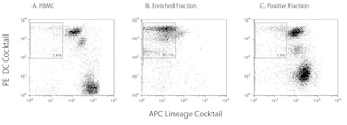
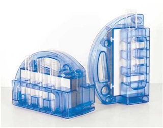
PBMCs are labeled with BD IMag™ anti-human CD11c Magnetic Particles - DM according to the following Magnetic Labeling Protocol. This labeled cell suspension is then placed within the magnetic field of the BD IMag™ Cell Separation Magnet (Cat. No. 552311). Labeled cells migrate toward the magnet (positive fraction), leaving the unlabeled cells in suspension so they can be drawn off (negative fraction). The tube is then removed from the magnetic field for resuspension of the positive fraction. The separation is repeated twice to increase the purity of the positive fraction. The magnetic separation steps are diagrammed in the Separation Flow Chart. After the positive fraction is washed, the small size of the magnetic particles allows the positive fraction to be further evaluated in downstream applications such as flow cytometry. This kit can be used in conjunction with the Human Dendritic Cell Enrichment Set - DM (Cat. No. 558420) to select for an enriched myeloid DC population (CD123 negative; CD11c positive) or plasmacytoid DC population (CD123 positive; CD11c negative).
MAGNETIC LABELING PROTOCOL
1. Prepare PBMCs from anti-coagulated human blood, preferably by density gradient centrifugation using Ficoll-Paque.™ Remove clumps of cells and/or debris by passing the suspension through a 70- m nylon cell strainer.
2. Dilute BD IMag™ Buffer (10X) (Cat. no. 552362) 1:10 with sterile distilled water or prepare 1X BD IMag™ buffer by supplementing Phosphate Buffered Saline with 0.5% BSA, 2 mM EDTA, and 0.09% sodium azide). Store at 4?C.
3. Wash cells with an excess volume of 1X BD IMag buffer, and carefully aspirate all the supernatant.
4. Vortex the BD IMag™ anti-human CD11c Particles - DM thoroughly, and add 50µl of particles for every 10 million total cells.
5. MIX THOROUGHLY. Incubate at room temperature for 30 minutes.
6. Bring the BD IMag-particle labeling volume up to 10 - 80 million cells/ml with 1X BD IMag™ buffer, and immediately place the tube on the BD IMag™ Cell Separation Magnet. Incubate for 8 - 10 minutes.
7. With the tube on the Cell Separation Magnet, carefully aspirate off the supernatant. This supernatant contains the negative fraction.
8. Remove the tube from the Cell Separation Magnet, and add 1 ml of 1X BD IMag buffer. Gently resuspend cells by pipetting up and down, and return the tube to the Cell Separation Magnet for another 2 - 4 minutes.
9. With the tube on the Cell Separation Magnet, carefully aspirate off the supernatant and discard.
10. Repeat Steps 8 and 9.
11. After the final wash step, resuspend the positive fraction in an appropriate buffer or media, and proceed with desired downstream application(s).
A more detailed protocol for the magnetic cell separation appears on the Technical Data Sheet accompanying the BD IMag™ Cell Separation Magnet (Cat. No. 552311).
The concentration of BD IMag™ anti-human CD11c Particles - DM suggested in the protocol has been optimized for the purification of CD11c-positive B leukocytes from human peripheral blood. When labeling target cell populations present at lower frequencies, fewer BD IMag™ particles can be used. Conversely, when labeling target cell populations that are present at higher frequencies, more particles should be used. To determine the optimal concentration of the BD IMag™ anti-human CD11c Particles - DM for a particular application, a titration in two-fold increments is recommended.
NOTE: Avoid nonspecific labeling by working quickly and keeping incubation times to a minimum.
Product Notices
- Caution: Sodium azide yields highly toxic hydrazoic acid under acidic conditions. Dilute azide compounds in running water before discarding to avoid accumulation of potentially explosive deposits in plumbing.
- Source of all serum proteins is from USDA inspected abattoirs located in the United States.
- BD IMag™ particles are prepared from carboxy-functionalized magnetic particles which are manufactured by Skold Technology and are licensed under US patent number 7,169,618.
- Ficoll-Paque is a trademark of Amersham Biosciences Limited.
- Please refer to www.bdbiosciences.com/us/s/resources for technical protocols.
Companion Products
BD IMag™ anti-human CD11c Particles - DM are magnetic nanoparticles that have monoclonal antibody conjugated to their surfaces. These particles are optimized for the positive selection or depletion of CD11c-bearing cells using the BD IMag™ Cell Separation Magnet. The CD11c antigen is present on monocytes and in low density on granulocytes and natural killer (NK) cells and dendritic cells (DC) in peripheral blood. It is also present on hairy-cell leukemias and on some acute myeloid leukemias (AML). CD11c reacts with an epitope on the α (150-kDa) chain of the CD11c/CD18 antigen complex. In addition, CD11c stains sinus histiocytes of lymph node, tonsil, and spleen; macrophages in the cortex and medulla of thymus; Kupffer cells in liver; elongated intertubular cells in kidney; and alveolar macrophages in lung tissue. In malignant tissue, the antibody stains true histiocytic lymphomas; it does not stain carcinomas or T or B lymphomas.
Development References (5)
-
Kurzinger K, Ho MK, Springer TA. Structural homology of a macrophage differentiation antigen and an antigen involved in T-cell-mediated killing. Nature. 1982; 296(5858):668-670. (Biology). View Reference
-
Lanier LL, Arnaout MA, Schwarting R, Warner NL, Ross GD. p150/95, Third member of the LFA-1/CR3 polypeptide family identified by anti-Leu M5 monoclonal antibody. Eur J Immunol. 1985; 15(7):713-718. (Biology). View Reference
-
Myones BL, Dalzell JG, Hogg N, Ross GD. Neutrophil and monocyte cell surface p150,95 has iC3b-receptor (CR4) activity resembling CR3.. J Clin Invest. 1988; 82(2):640-51. (Biology). View Reference
-
Sanchez-Madrid F, Simon P, Thompson S, Springer TA. Mapping of antigenic and functional epitopes on the alpha- and beta-subunits of two related mouse glycoproteins involved in cell interactions, LFA-1 and Mac-1. J Exp Med. 1983; 158(2):586-602. (Biology). View Reference
-
Schwarting R, Stein H, Wang CY. The monoclonal antibodies alpha S-HCL 1 (alpha Leu-14) and alpha S-HCL 3 (alpha Leu-M5) allow the diagnosis of hairy cell leukemia. Blood. 1985; 65(4):974-983. (Clone-specific). View Reference
Please refer to Support Documents for Quality Certificates
Global - Refer to manufacturer's instructions for use and related User Manuals and Technical data sheets before using this products as described
Comparisons, where applicable, are made against older BD Technology, manual methods or are general performance claims. Comparisons are not made against non-BD technologies, unless otherwise noted.
For Research Use Only. Not for use in diagnostic or therapeutic procedures.