-
Reagents
- Flow Cytometry Reagents
-
Western Blotting and Molecular Reagents
- Immunoassay Reagents
-
Single-Cell Multiomics Reagents
- BD® OMICS-Guard Sample Preservation Buffer
- BD® AbSeq Assay
- BD® Single-Cell Multiplexing Kit
- BD Rhapsody™ ATAC-Seq Assays
- BD Rhapsody™ Whole Transcriptome Analysis (WTA) Amplification Kit
- BD Rhapsody™ TCR/BCR Next Multiomic Assays
- BD Rhapsody™ Targeted mRNA Kits
- BD Rhapsody™ Accessory Kits
- BD® OMICS-One Protein Panels
-
Functional Assays
-
Microscopy and Imaging Reagents
-
Cell Preparation and Separation Reagents
-
- BD® OMICS-Guard Sample Preservation Buffer
- BD® AbSeq Assay
- BD® Single-Cell Multiplexing Kit
- BD Rhapsody™ ATAC-Seq Assays
- BD Rhapsody™ Whole Transcriptome Analysis (WTA) Amplification Kit
- BD Rhapsody™ TCR/BCR Next Multiomic Assays
- BD Rhapsody™ Targeted mRNA Kits
- BD Rhapsody™ Accessory Kits
- BD® OMICS-One Protein Panels
- France (English)
-
Change country/language
Old Browser
This page has been recently translated and is available in French now.
Looks like you're visiting us from United States.
Would you like to stay on the current country site or be switched to your country?
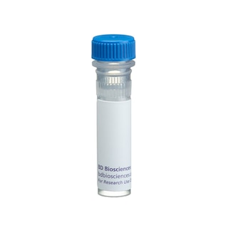
BD Transduction Laboratories™ Purified Mouse Anti-Mouse Thrombospondin-2
Clone 4/Thrombospondin-2 (RUO)


Western blot analysis of Thrombospondin-2 on a RSV-3T3 cell lysate. Lane 1: 1:250, lane 2: 1:500, lane 3: 1:1000 dilution of the mouse anti-mouse thrombospondin-2 antibody.


BD Transduction Laboratories™ Purified Mouse Anti-Mouse Thrombospondin-2

Regulatory Status Legend
Any use of products other than the permitted use without the express written authorization of Becton, Dickinson and Company is strictly prohibited.
Preparation And Storage
Recommended Assay Procedures
Western blot: Please refer to http://www.bdbiosciences.com/pharmingen/protocols/Western_Blotting.shtml
Product Notices
- Since applications vary, each investigator should titrate the reagent to obtain optimal results.
- Please refer to www.bdbiosciences.com/us/s/resources for technical protocols.
- Caution: Sodium azide yields highly toxic hydrazoic acid under acidic conditions. Dilute azide compounds in running water before discarding to avoid accumulation of potentially explosive deposits in plumbing.
- Source of all serum proteins is from USDA inspected abattoirs located in the United States.
Data Sheets
Companion Products

.png?imwidth=320)
Cell adhesion interactions involve large extracellular glycoproteins such as fibronectin, laminins, and thrombospondins. The thrombospondins (TSPs), a small family of secreted glycoproteins, can be separated into two subfamilies based on structural similarity. TSP-1 and TSP-2 are homotrimers of monomeric 145 kDa chains, while TSP-3,4 and 5 are homopentamers of lower MW chains. TSP-1 and TSP-2 interact with several of the same cell surface receptors and have been termed matricellular proteins. However, the functional roles of TSP-1 and TSP-2 are poorly understood. Study of TSP-2 deficient mice has shed some light on the functional role of this protein. Although these mice appear normal, they in fact exhibit a wide variety of irregularities. These include abnormal collagen fiber development which results in fragile skin and highly flexible tendons and ligaments. They also exhibit increased cortical thickness in long bones and abnormal bleeding time. These results, together with the developmental expression pattern of TSP-2, suggest that it functions as a modulator of the cell surface properties of mesenchymal cells which, in turn, affects cell functions such as adhesion and migration.
This antibody is routinely tested by western blot analysis. Otehr applications were tested at BD Biosciences Pharmingen during antibody development only or reported in the literature.
Development References (3)
-
Kyriakides TR, Zhu YH, Smith LT, et al. Mice that lack thrombospondin 2 display connective tissue abnormalities that are associated with disordered collagen fibrillogenesis, an increased vascular density, and a bleeding diathesis. J Cell Biol. 1998; 140(2):419-430. (Biology). View Reference
-
Kyriakides TR, Zhu YH, Yang Z, Bornstein P. The distribution of the matricellular protein thrombospondin 2 in tissues of embryonic and adult mice. J Histochem Cytochem. 1998; 46(9):1007-1015. (Biology). View Reference
-
LaBell TL, Milewicz DJ, Disteche CM, Byers PH. Thrombospondin II: partial cDNA sequence, chromosome location, and expression of a second member of the thrombospondin gene family in humans. Genomics. 1992; 12(3):421-429. (Biology). View Reference
Please refer to Support Documents for Quality Certificates
Global - Refer to manufacturer's instructions for use and related User Manuals and Technical data sheets before using this products as described
Comparisons, where applicable, are made against older BD Technology, manual methods or are general performance claims. Comparisons are not made against non-BD technologies, unless otherwise noted.
For Research Use Only. Not for use in diagnostic or therapeutic procedures.