







Preparation And Storage


Recommended Assay Procedures
Chemokine receptors are know to internalize during manipulation resulting in low frequency expression. Immunophenotyping studies of chemokine receptors need to be performed on freshly collected whole blood (<24 Hrs). Incubation with the antibody should be done in the dark. Cellular manipulation, such as Ficoll separation, freezing, or exposure to cold temperatures prior to staining have been shown to cause a decrease in staining intensity and inconsistent results.
The purified 3D12 antibody can be used for the immunofluorescent staining and flow cytometric analyses of human leukocytes and cell lines that express CCR7 (see Figure). A multiple-step staining procedure is strongly recommended to amplify immunofluorescent signals for the flow cytometric analysis of human CCR7 expression:
1. Incubate 10e6 cells with 0.1 - 0.5 µg of Purified Rat Anti-Human CCR7 (CD197) at 4°C for 15 - 20 minutes. Wash cells two times with staining medium containing sodium azide (e.g., Dulbecco's PBS or tissue culture medium [without phenol red and biotin] with 0.09% sodium azide and 2% heat-inactivated FCS or 0.2% BSA).
2. Incubate the cells with 0.25 µg of Biotin Mouse Anti-Rat IgG2a (Cat. No. 553894) at 4°C for 20 minutes. Wash cells two times.
3. Incubate the cells with ≤ 0.06 µg of streptavidin-phycoerythrin (Cat. No. 554061) at 4°C for 20 minutes. Wash two times. Resuspend cells instaining medium and analyze stained cells with a FACScan™ Flow Cytometer (BDIS, San Jose, CA) using appropriate specificity and compensation controls.
Product Notices
- Since applications vary, each investigator should titrate the reagent to obtain optimal results.
- An isotype control should be used at the same concentration as the antibody of interest.
- Caution: Sodium azide yields highly toxic hydrazoic acid under acidic conditions. Dilute azide compounds in running water before discarding to avoid accumulation of potentially explosive deposits in plumbing.
- Sodium azide is a reversible inhibitor of oxidative metabolism; therefore, antibody preparations containing this preservative agent must not be used in cell cultures nor injected into animals. Sodium azide may be removed by washing stained cells or plate-bound antibody or dialyzing soluble antibody in sodium azide-free buffer. Since endotoxin may also affect the results of functional studies, we recommend the NA/LE (No Azide/Low Endotoxin) antibody format, if available, for in vitro and in vivo use.
- For fluorochrome spectra and suitable instrument settings, please refer to our Multicolor Flow Cytometry web page at www.bdbiosciences.com/colors.
- Please refer to www.bdbiosciences.com/us/s/resources for technical protocols.
Companion Products



.png?imwidth=320)
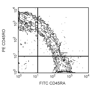
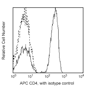
The monoclonal antibody 3D12 reacts with the human CC chemokine receptor, CCR7. CCR7 (previously known as BLR-2, EBI-1 and CMKBR7), a seven-transmembrane, G-protein-coupled receptor, is the specific receptor for CC chemokines, MIP-3β/Exodus 3/ELC/ CCL19 and 6Ckine/Exodus 2/SLC/TCA4/CCL21. It has been shown that CCR7 mRNA is expressed mainly in lymphoid tissues including spleen, lymph nodes and tonsil. CCR7 mRNA was also detected in peripheral T and B lymphocytes, in bone marrow and cord blood CD34-positive cells and mature dendritic cells. The human CCR7 gene, unlike other CC chemokine receptor genes, has been mapped to chromosome 17q12. The immunogen used to generate 3D12 hybridoma was the N-terminus as well as parts of the second extracellular loop of human CCR7 protein. The monoclonal antibody 3D12 recognizes an epitope mapping to the N-terminus of human CCR7.
Development References (15)
-
Birkenbach M, Josefsen K, Yalamanchili R, Lenoir G, Kieff E. Epstein-Barr virus-induced genes: first lymphocyte-specific G protein-coupled peptide receptors. Nature. 1993; 67(4):2209-2220. (Biology). View Reference
-
Britschgi MR, Link A, Lissandrin TK, Luther SA. Dynamic modulation of CCR7 expression and function on naive T lymphocytes in vivo. J Immunol. 2008; 181(11):7681-7688. (Biology). View Reference
-
Burgstahler R, Kempkes B, Steube K, Lipp M. Expression of the chemokine receptor BLR2/EBI1 is specifically transactivated by Epstein-Barr virus nuclear antigen 2. Biochem Biophys Res Commun. 1995; 215(2):737-743. (Biology). View Reference
-
Forster R, Davalos-Misslitz AC, Rot A. CCR7 and its ligands: balancing immunity and tolerance. Nat Rev Immunol. 2008; 8(5):362-371. (Biology). View Reference
-
Kim CH, Pelus LM, White JR, Broxmeyer HE. Macrophage-inflammatory protein-3 beta/EBI1-ligand chemokine/CK beta-11, a CC chemokine, is a chemoattractant with a specificity for macrophage progenitors among myeloid progenitor cells. J Immunol. 1998; 161(5):2580-2585. (Biology). View Reference
-
Kurobe, H., Liu, et al. CCR7-dependent cortex-to-medulla migration of positively selected thymocytes is essential for establishing central tolerance. Immunity. 2006; 24(2):165-177. (Biology). View Reference
-
Lipp M, Burgstahler R, Muller G, et al. Functional organization of secondary lymphoid organs by the chemokine system. Curr Top Microbiol Immunol. 2000; 251:173-179. (Clone-specific). View Reference
-
Ohl L, Mohaupt M, Czeloth N, et al. CCR7 governs skin dendritic cell migration under inflammatory and steady-state conditions. Immunity. 2004; 21(2):279-288. (Biology). View Reference
-
Ritter U, Wiede F, Mielenz D, Kiafard Z, Zwirner J, Korner H. Analysis of the CCR7 expression on murine bone marrow-derived and spleen dendritic cells.. J Leukoc Biol. 2004; 76(2):472-476. (Biology). View Reference
-
Sallusto F, Lenig D, Forster R, Lipp M, Lanzavecchia A. Two subsets of memory T lymphocytes with distinct homing potentials and effector functions. Nature. 1999; 401(6754):708-712. (Clone-specific). View Reference
-
Schweickart VL, Raport CJ, Godiska R, et al. Cloning of human and mouse EBI1, a lymphoid-specific G-protein-coupled receptor encoded on human chromosome 17q12-q21.2. Genomics. 1994; 23(3):643-650. (Biology). View Reference
-
Yanagihara S, Komura E, Nagafune J, Watarai H, Yamaguchi Y. EBI1/CCR7 is a new member of dendritic cell chemokine receptor that is up-regulated upon maturation. J Immunol. 1998; 161(6):3096-3102. (Biology). View Reference
-
Yoshida R, Imai T, Hieshima K, et al. Molecular cloning of a novel human CC chemokine EBI1-ligand chemokine that is a specific functional ligand for EBI1, CCR7. J Biol Chem. 1997; 272(21):13803-13809. (Biology). View Reference
-
Yoshida R, Nagira M, Imai T, et al. EBI1-ligand chemokine (ELC) attracts a broad spectrum of lymphocytes: activated T cells strongly up-regulate CCR7 and efficiently migrate toward ELC. Int Immunol. 1998; 10(7):901-910. (Biology). View Reference
-
Yoshida R, Nagira M, Kitaura M, Imagawa N, Imai T, Yoshie O. Secondary lymphoid-tissue chemokine is a functional ligand for the CC chemokine receptor CCR7. J Biol Chem. 1998; 273(12):7118-7122. (Biology). View Reference
Please refer to Support Documents for Quality Certificates
Global - Refer to manufacturer's instructions for use and related User Manuals and Technical data sheets before using this products as described
Comparisons, where applicable, are made against older BD Technology, manual methods or are general performance claims. Comparisons are not made against non-BD technologies, unless otherwise noted.
For Research Use Only. Not for use in diagnostic or therapeutic procedures.
