Old Browser
This page has been recently translated and is available in French now.
Looks like you're visiting us from {countryName}.
Would you like to stay on the current country site or be switched to your country?


Regulatory Status Legend
Any use of products other than the permitted use without the express written authorization of Becton, Dickinson and Company is strictly prohibited.
Preparation And Storage
Recommended Assay Procedures
For optimal and reproducible results, BD Horizon Brilliant Stain Buffer should be used anytime two or more BD Horizon Brilliant dyes (including BD OptiBuild Brilliant reagents) are used in the same experiment. Fluorescent dye interactions may cause staining artifacts which may affect data interpretation. The BD Horizon Brilliant Stain Buffer was designed to minimize these interactions. More information can be found in the Technical Data Sheet of the BD Horizon Brilliant Stain Buffer (Cat. No. 563794).
Product Notices
- This antibody was developed for use in flow cytometry.
- The production process underwent stringent testing and validation to assure that it generates a high-quality conjugate with consistent performance and specific binding activity. However, verification testing has not been performed on all conjugate lots.
- Researchers should determine the optimal concentration of this reagent for their individual applications.
- An isotype control should be used at the same concentration as the antibody of interest.
- Caution: Sodium azide yields highly toxic hydrazoic acid under acidic conditions. Dilute azide compounds in running water before discarding to avoid accumulation of potentially explosive deposits in plumbing.
- For fluorochrome spectra and suitable instrument settings, please refer to our Multicolor Flow Cytometry web page at www.bdbiosciences.com/colors.
- Please refer to www.bdbiosciences.com/us/s/resources for technical protocols.
- BD Horizon Brilliant Stain Buffer is covered by one or more of the following US patents: 8,110,673; 8,158,444; 8,575,303; 8,354,239.
- BD Horizon Brilliant Ultraviolet 395 is covered by one or more of the following US patents: 8,158,444; 8,575,303; 8,354,239.
Companion Products


The MEC13.3 antibody specifically recognizes CD31, also known as PECAM-1 (Platelet Endothelial Cell Adhesion Molecule-1). CD31 is a 130 kDa integral membrane protein, a member of the immunoglobulin superfamily, that mediates cell-to-cell adhesion. CD31 is expressed constitutively on the surface of adult and embryonic endothelial cells and is also expressed on many peripheral leukocytes and platelets. It has also been detected on bone marrow-derived hematopoietic stem cells and embryonic stem cells. CD31 is involved in the transendothelial emigration of neutrophils, and neutrophil PECAM-1 appears to be down-regulated after extravasation into inflamed tissues. Multiple alternatively spliced isoforms are detected during early post-implantation embryonic development; this alternative splicing is involved in the regulation of ligand specificity. CD38 and vitronectin receptor (αvβ3 integrin, CD51/CD61) are proposed to be ligands for CD31. CD31-mediated endothelial cell-cell interactions are involved in angiogenesis. The MEC13.3 mAb inhibits a variety of in vitro and in vivo functions mediated by CD31.
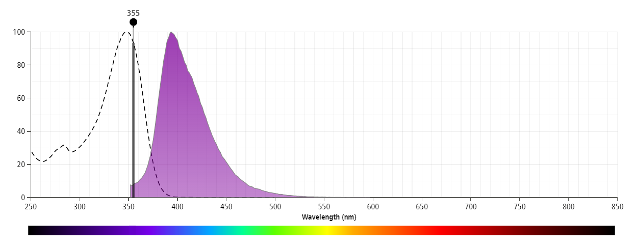
The antibody was conjugated to BD Horizon™ BUV395 which is part of the BD Horizon Brilliant™ Ultraviolet family of dyes. This dye has been exclusively developed by BD Biosciences to have minimal spillover into other detectors, making it an optimal choice for multicolor flow cytometry. With an Ex Max at 348 nm and an Em Max at 395 nm, BD Horizon BUV395 can be excited with a 355 nm laser and detected with a 379/28 filter.

Development References (13)
-
Baldwin HS, Shen HM, Yan HC, et al. Platelet endothelial cell adhesion molecule-1 (PECAM-1/CD31): alternatively spliced, functionally distinct isoforms expressed during mammalian cardiovascular development. Development. 1994; 120(9):2539-2953. (Clone-specific: Blocking, Flow cytometry, Immunohistochemistry). View Reference
-
Christofidou-Solomidou M, Nakada MT, Williams J, Muller WA, DeLisser HM. Neutrophil platelet endothelial cell adhesion molecule-1 participates in neutrophil recruitment at inflammatory sites and is down-regulated after leukocyte extravasation. J Immunol. 1997; 158(10):4872-4878. (Clone-specific: Flow cytometry, Inhibition, In vivo exacerbation). View Reference
-
DeLisser HM, Christofidou-Solomidou M, Strieter RM, et al. Involvement of endothelial PECAM-1/CD31 in angiogenesis. Am J Pathol. 1997; 151(3):671-677. (Clone-specific: Inhibition, In vivo exacerbation). View Reference
-
DeLisser HM, Newman PJ, Albelda SM. Molecular and functional aspects of PECAM-1/CD31. Immunol Today. 1994; 15(10):490-495. (Biology). View Reference
-
Duncan GS, Andrew DP, Takimoto H, et al. Genetic evidence for functional redundancy of Platelet/Endothelial cell adhesion molecule-1 (PECAM-1): CD31-deficient mice reveal PECAM-1-dependent and PECAM-1-independent functions. J Immunol. 1999; 162(5):3022-3030. (Clone-specific: Flow cytometry, Immunohistochemistry). View Reference
-
Famiglietti J, Sun J, DeLisser HM, Albelda SM. Tyrosine residue in exon 14 of the cytoplasmic domain of platelet endothelial cell adhesion molecule-1 (PECAM-1/CD31) regulates ligand binding specificity. J Cell Biol. 1997; 138(6):1425-1435. (Biology). View Reference
-
Horenstein AL, Stockinger H, Imhof BA, Malavasi F. CD38 binding to human myeloid cells is mediated by mouse and human CD31. Biochem J. 1998; 330(3):1129-1135. (Biology). View Reference
-
Ling V, Luxenberg D, Wang J, et al. Structural identification of the hematopoietic progenitor antigen ER-MP12 as the vascular endothelial adhesion molecule PECAM-1 (CD31). Eur J Immunol. 1997; 27(2):509-514. (Clone-specific: Flow cytometry). View Reference
-
Piali L, Hammel P, Uherek C, et al. CD31/PECAM-1 is a ligand for alpha v beta 3 integrin involved in adhesion of leukocytes to endothelium. J Cell Biol. 1995; 130(2):451-460. (Biology). View Reference
-
Rosenblum WI, Murata S, Nelson GH, Werner PK, Ranken R, Harmon RC. Anti-CD31 delays platelet adhesion/aggregation at sites of endothelial injury in mouse cerebral arterioles. Am J Pathol. 1994; 145(1):33-36. (Clone-specific: Flow cytometry, Immunohistochemistry, Immunoprecipitation, In vivo exacerbation). View Reference
-
Suri C, Jones PF, Patan S, et al. Requisite role of angiopoietin-1, a ligand for the TIE2 receptor, during embryonic angiogenesis. Cell. 1996; 87(7):1171-1180. (Clone-specific: Immunohistochemistry). View Reference
-
Vanzulli S, Gazzaniga S, Braidot MF, et al. Detection of endothelial cells by MEC 13.3 monoclonal antibody in mice mammary tumors. Biocell. 1997; 21(1):39-46. (Clone-specific: Immunohistochemistry). View Reference
-
Vecchi A, Garlanda C, Lampugnani MG, et al. Monoclonal antibodies specific for endothelial cells of mouse blood vessels. Their application in the identification of adult and embryonic endothelium. Eur J Cell Biol. 1994; 63(2):247-254. (Immunogen: ELISA, Flow cytometry, Fluorescence microscopy, Immunofluorescence, Immunohistochemistry, Immunoprecipitation). View Reference
Please refer to Support Documents for Quality Certificates
Global - Refer to manufacturer's instructions for use and related User Manuals and Technical data sheets before using this products as described
Comparisons, where applicable, are made against older BD Technology, manual methods or are general performance claims. Comparisons are not made against non-BD technologies, unless otherwise noted.
For Research Use Only. Not for use in diagnostic or therapeutic procedures.
Report a Site Issue
This form is intended to help us improve our website experience. For other support, please visit our Contact Us page.