Old Browser
This page has been recently translated and is available in French now.
Looks like you're visiting us from {countryName}.
Would you like to stay on the current country site or be switched to your country?


Western blot analysis of eNOS/NOS Type III on human endothelial cell lysate. Lane 1: 1:2500, lane 2: 1:5000, lane 3: 1:10000 dilution of anti-eNOS/NOS Type III.

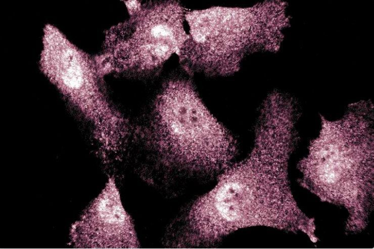
Immunofluorescent staining of Endothelial Cells


BD Transduction Laboratories™ Purified Mouse Anti-eNOS/NOS Type III

BD Transduction Laboratories™ Purified Mouse Anti-eNOS/NOS Type III

Regulatory Status Legend
Any use of products other than the permitted use without the express written authorization of Becton, Dickinson and Company is strictly prohibited.
Preparation And Storage
Product Notices
- Since applications vary, each investigator should titrate the reagent to obtain optimal results.
- Please refer to www.bdbiosciences.com/us/s/resources for technical protocols.
- Caution: Sodium azide yields highly toxic hydrazoic acid under acidic conditions. Dilute azide compounds in running water before discarding to avoid accumulation of potentially explosive deposits in plumbing.
- Source of all serum proteins is from USDA inspected abattoirs located in the United States.
- For fluorochrome spectra and suitable instrument settings, please refer to our Multicolor Flow Cytometry web page at www.bdbiosciences.com/colors.
Nitric oxide synthase (NOS), a cell-type specific enzyme, catalyzes the synthesis of nitric oxide (NO). NO is a short-lived radical that transmits signals involved in vasorelaxation, neurotransmission, and cytotoxicity. In neurons and endothelial cells, constitutive NOS (cNOS) is activated by agonists that increase intracellular Ca[2+] levels and enhance calmodulin binding. Neuronal NOS (mNOS) and endothelial NOS (eNOS) have recognition sites for NADPH, FAD, FMN, and calmodulin and both are regulated in a similar manner. The human forms exhibit 52% amino acid identity. However, they are distinct gene products of about 155 kDa (mNOS) and 140 kDA (eNOS). The eNOS gene was cloned from human vascular endothelium as well as from bovine aortic endothelial cells (BAEC). eNOS protein has a unique N-myristylation consensus sequence that may explain its membrane localization.
Investigators should note that clone 3/eNOS/NOS Type III was never immunohistochemistry (IHC) tested on rat tissues while in development.
Development References (5)
-
Cao S, Yao J, McCabe TJ, et al. Direct interaction between endothelial nitric-oxide synthase and dynamin-2. Implications for nitric-oxide synthase function. J Biol Chem. 2001; 276(17):14249-14256. (Clone-specific: Immunofluorescence, Immunoprecipitation, Western blot). View Reference
-
Kincer JF, Uittenbogaard A, Dressman J, et al. Hypercholesterolemia promotes a CD36-dependent and endothelial nitric-oxide synthase-mediated vascular dysfunction. J Biol Chem. 2002; 277(26):23525-23533. (Clone-specific: Western blot). View Reference
-
Laufs U, Endres M, Stagliano N, et al. Neuroprotection mediated by changes in the endothelial actin cytoskeleton. J Clin Invest. 2000; 106(1):15-24. (Clone-specific: Immunofluorescence, Immunohistochemistry). View Reference
-
Thomas SR, Chen K, Keaney JF Jr. Hydrogen peroxide activates endothelial nitric-oxide synthase through coordinated phosphorylation and dephosphorylation via a phosphoinositide 3-kinase-dependent signaling pathway. J Biol Chem. 2002; 277(8):6017-6024. (Clone-specific: Western blot). View Reference
-
Zhao H, Dugas N, Mathiot C, et al. B-cell chronic lymphocytic leukemia cells express a functional inducible nitric oxide synthase displaying anti-apoptotic activity. Blood. 1998; 92(3):1031-1043. (Clone-specific: Flow cytometry). View Reference
Please refer to Support Documents for Quality Certificates
Global - Refer to manufacturer's instructions for use and related User Manuals and Technical data sheets before using this products as described
Comparisons, where applicable, are made against older BD Technology, manual methods or are general performance claims. Comparisons are not made against non-BD technologies, unless otherwise noted.
For Research Use Only. Not for use in diagnostic or therapeutic procedures.
Report a Site Issue
This form is intended to help us improve our website experience. For other support, please visit our Contact Us page.