-
Reagents
- Flow Cytometry Reagents
-
Western Blotting and Molecular Reagents
- Immunoassay Reagents
-
Single-Cell Multiomics Reagents
- BD® OMICS-Guard Sample Preservation Buffer
- BD® AbSeq Assay
- BD® Single-Cell Multiplexing Kit
- BD Rhapsody™ ATAC-Seq Assays
- BD Rhapsody™ Whole Transcriptome Analysis (WTA) Amplification Kit
- BD Rhapsody™ TCR/BCR Next Multiomic Assays
- BD Rhapsody™ Targeted mRNA Kits
- BD Rhapsody™ Accessory Kits
- BD® OMICS-One Protein Panels
-
Functional Assays
-
Microscopy and Imaging Reagents
-
Cell Preparation and Separation Reagents
-
- BD® OMICS-Guard Sample Preservation Buffer
- BD® AbSeq Assay
- BD® Single-Cell Multiplexing Kit
- BD Rhapsody™ ATAC-Seq Assays
- BD Rhapsody™ Whole Transcriptome Analysis (WTA) Amplification Kit
- BD Rhapsody™ TCR/BCR Next Multiomic Assays
- BD Rhapsody™ Targeted mRNA Kits
- BD Rhapsody™ Accessory Kits
- BD® OMICS-One Protein Panels
- France (English)
-
Change country/language
Old Browser
This page has been recently translated and is available in French now.
Looks like you're visiting us from United States.
Would you like to stay on the current country site or be switched to your country?
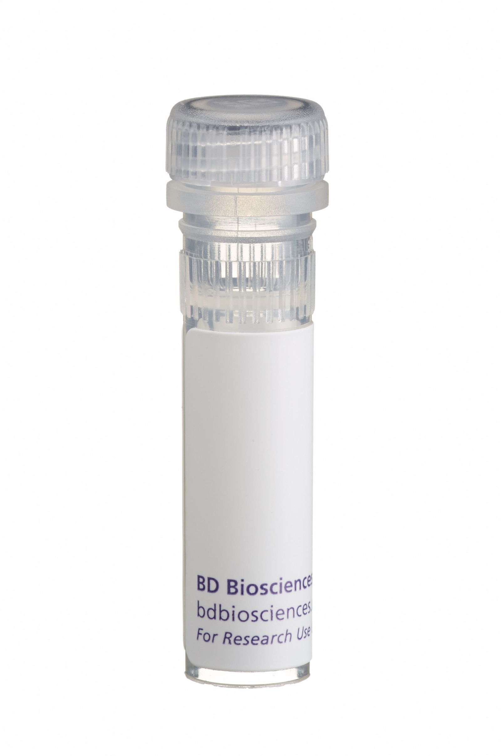
BD Pharmingen™ Recombinant Mouse GM-CSF


Regulatory Status Legend
Any use of products other than the permitted use without the express written authorization of Becton, Dickinson and Company is strictly prohibited.
Product Details
Description
Mouse granulocyte-macrophage colony-stimulating factor (GM-CSF) is a cytokine made by activated T cells, macrophages, vascular endothelial cells and fibroblasts. It has potent stimulatory effects on the growth and differentiation of bone marrow progenitor cells that generate granulocytes, monocytes/macrophages, and megakaryocytes. In peripheral tissues, GM-CSF can act on mature leukocytes to promote inflammatory responses. Mouse GM-CSF runs as a 15.5 - 19 kD band in reducing and non-reducing SDS-PAGE analysis. Mouse GM-CSF protein contains 124 amino acid residues. Recombinat mouse GM-CSF (Cat. No. 554586) is supplied as a frozen liquid comprised of 0.22 um sterile-filtered aqueous buffered solution containing bovine serum albumin, with no preservatives. Recombinant mouse GM-CSF is ≥ 95% pure as determined by SDS-PAGE and an absorbance assay based on the Beers-Lambert law. The endotoxin level is ≤ 0.1 ng per µg of mouse GM-CSF, as measured in chromogenic LAL assay.
Preparation And Storage
Recommended Assay Procedures
Upon initial thawing, recombinant mouse GM-CSF (Cat. No. 554586) should be aliquoted into polypropylene microtubes and frozen at -80°C for future use. Alternatively, the product can be diluted in sterile neutral buffer containing not less than 0.5 - 10 mg/mL carrier protein, such as human or bovine albumin, aliquoted and stored at -80°C. For in vitro biological assay use, carrier protein concentrations of 0.5 - 1 mg/mL are recommended. For use as an ELISA standard, carrier-protein concentrations of 5 - 10 mg/mL are recommended. Failure to add carrier protein or store at indicated temperatures may result in a loss of activity. This product should not be diluted to less than 5 μg/mL for long term storage. Carrier proteins should be pre-screened for possible effects in each investigator's experimental system. Carrier proteins may have an undesired influence on experimental results due to toxicity, high endotoxin levels or possible blocking activity.
ELISA Standard: Recombinant mouse GM-CSF (Cat. No. 554586) can be useful as a quantitative standard for measuring mouse GM-CSF protein levels using sandwich ELISA with the purified MP1-22E9 antibody (Cat. No. 554404) as a capture antibody and biotinylated MP1-31G6 antibody (Cat. No. 554407) as the detection antibody. To obtain linear standard curves, investigators may want to consider using doubling dilutions of recombinant mouse GM-CSF standard from 2,000 - 15 pg/mL to be included in each ELISA plate. For measuring mouse GM-CSF in serum or plasma, investigators are highly encouraged to use the BD OptEIA™ Mouse GM-CSF ELISA Set (Cat. No. 555167).
Bioassay: Investigators are advised that the Bioassay application is not routinely tested for this material and are highly encouraged to both titrate this material and include appropriate controls in relevant experiments. An activity range of 0.25 - 5.0 x 10^9 units/mg, encompassing an
ED50= 2 - 35 pg/mL, has previously been reported using MC/9 as indicator cells for proliferation, with a unit defined as the amount of material needed to stimulate a half-maximal response at cytokine saturation.
Blocking: Recombinant mouse GM-CSF (Cat. No. 554586) can be useful as a blocking control for flow cytometric analysis when used with PE-conjugated MP1-22E9 antibody (Cat. No. 554406). Investigators are advised that the blocking application is not routinely tested for this material. Intracellular cytokine staining techniques and the use of blocking controls are described in detail by C. Prussin and D. Metcalfe.
Product Notices
- Since applications vary, each investigator should titrate the reagent to obtain optimal results.
- Please refer to www.bdbiosciences.com/us/s/resources for technical protocols.
- Source of all serum proteins is from USDA inspected abattoirs located in the United States.
Data Sheets
Companion Products


Development References (6)
-
Gasson JC. Molecular physiology of granulocyte-macrophage colony-stimulating factor. Blood. 1991; 77(6):1131-1145. (Biology). View Reference
-
Gough NM, Gough J, Metcalf D, et al. Molecular cloning of cDNA encoding a murine haematopoietic growth regulator, granulocyte-macrophage colony stimulating factor. Nature. 1984; 309(5971):763-767. (Biology). View Reference
-
Miyatake S, Otsuka T, Yokota T, Lee F, Arai K. Structure of the chromosomal gene for granulocyte-macrophage colony stimulating factor: comparison of the mouse and human genes. EMBO J. 1985; 4(10):2561-2568. (Biology). View Reference
-
Price V, Mochizuki D, March CJ, et al. Expression, purification and characterization of recombinant murine granulocyte-macrophage colony-stimulating factor and bovine interleukin-2 from yeast. Gene. 1987; 55:287-293. (Biology). View Reference
-
Prussin C, Metcalfe DD. Detection of intracytoplasmic cytokine using flow cytometry and directly conjugated anti-cytokine antibodies. J Immunol Methods. 1995; 188(1):117-128. (Methodology: Flow cytometry). View Reference
-
Thompson-Snipes L, Dhar V, Bond MW, Mosmann TR, Moore KW, Rennick DM. Interleukin 10: a novel stimulatory factor for mast cells and their progenitors. J Exp Med. 1991; 173(2):507-510. (Methodology). View Reference
Please refer to Support Documents for Quality Certificates
Global - Refer to manufacturer's instructions for use and related User Manuals and Technical data sheets before using this products as described
Comparisons, where applicable, are made against older BD Technology, manual methods or are general performance claims. Comparisons are not made against non-BD technologies, unless otherwise noted.
For Research Use Only. Not for use in diagnostic or therapeutic procedures.