Old Browser
This page has been recently translated and is available in French now.
Looks like you're visiting us from {countryName}.
Would you like to stay on the current country site or be switched to your country?


Profile of resting peripheral blood lymphocytes analyzed by flow cytometry

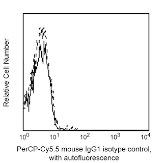
Multivariate flow cytometric analysis of CD38 expression on human peripheral blood leucocyte populations. Human whole blood was stained with either PerCP-Cy5.5 Mouse IgG1 κ Isotype Control (Cat. No. 550795; Left Plot) or PerCP-Cy5.5 Mouse Anti-Human CD38 antibody (Cat. No. 551400/561106; Right Plot) at 0.5 µg/test. Erythrocytes were lysed with BD Pharm Lyse™ Lysing Buffer (Cat. No. 555899). The bivariate pseudocolor density plot showing CD38 expression (or Ig Isotype control staining) versus side light-scatter (SSC-A) signals were derived from events with the forward and side light-scatter characteristics of intact leucocytes. Flow cytometry and data analysis were performed using a BD™ LSR II Flow Cytometer System and FlowJo™ software. Data shown on this Technical Data Sheet are not lot specific.


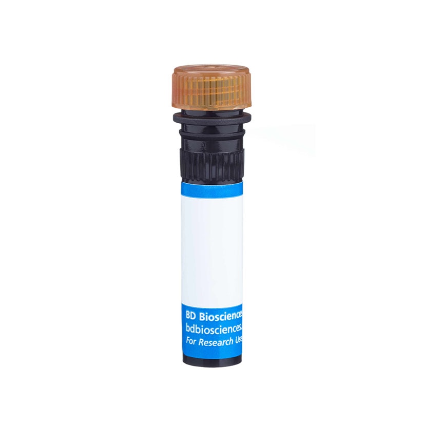
BD Pharmingen™ PerCP-Cy™5.5 Mouse Anti-Human CD38

BD Pharmingen™ PerCP-Cy™5.5 Mouse Anti-Human CD38

Regulatory Status Legend
Any use of products other than the permitted use without the express written authorization of Becton, Dickinson and Company is strictly prohibited.
Preparation And Storage
Recommended Assay Procedures
BD® CompBeads can be used as surrogates to assess fluorescence spillover (Compensation). When fluorochrome conjugated antibodies are bound to BD® CompBeads, they have spectral properties very similar to cells. However, for some fluorochromes there can be small differences in spectral emissions compared to cells, resulting in spillover values that differ when compared to biological controls. It is strongly recommended that when using a reagent for the first time, users compare the spillover on cells and BD CompBeads to ensure that BD® CompBeads are appropriate for your specific cellular application.
Product Notices
- Since applications vary, each investigator should titrate the reagent to obtain optimal results.
- An isotype control should be used at the same concentration as the antibody of interest.
- Caution: Sodium azide yields highly toxic hydrazoic acid under acidic conditions. Dilute azide compounds in running water before discarding to avoid accumulation of potentially explosive deposits in plumbing.
- Please refer to www.bdbiosciences.com/us/s/resources for technical protocols.
- For fluorochrome spectra and suitable instrument settings, please refer to our Multicolor Flow Cytometry web page at www.bdbiosciences.com/colors.
- PerCP-Cy5.5–labelled antibodies can be used with FITC- and R-PE–labelled reagents in single-laser flow cytometers with no significant spectral overlap of PerCP-Cy5.5, FITC, and R-PE fluorescence.
- PerCP-Cy5.5 is optimized for use with a single argon ion laser emitting 488-nm light. Because of the broad absorption spectrum of the tandem fluorochrome, extra care must be taken when using dual-laser cytometers, which may directly excite both PerCP and Cy5.5™. We recommend the use of cross-beam compensation during data acquisition or software compensation during data analysis.
- Please observe the following precautions: Absorption of visible light can significantly alter the energy transfer occurring in any tandem fluorochrome conjugate; therefore, we recommend that special precautions be taken (such as wrapping vials, tubes, or racks in aluminum foil) to prevent exposure of conjugated reagents, including cells stained with those reagents, to room illumination.
- Please refer to http://regdocs.bd.com to access safety data sheets (SDS).
- Cy is a trademark of Global Life Sciences Solutions Germany GmbH or an affiliate doing business as Cytiva.
The HIT2 monoclonal antibody specifically binds to CD38. The CD38 antigen is also known as T10, ADP-ribosyl cyclase 1, and cyclic ADP ribose hydrolase 1. CD38 is a 45 kDa type II single-chain transmembrane glycoprotein present on thymocytes, activated T cells and terminally differentiated B cells (plasma cells). CD38 is expressed by other cells including monocytes, macrophages, dendritic cells, NK cells, myeloid and erythroid precursors and some epithelial cells. The CD38 antigen acts as an ectoenzyme that catalyzes the synthesis and hydrolysis of a Ca++ mobilizing agent, cyclic ADP-ribose. This intracellular calcium plays an important role in cell signaling pathways leading to cellular growth, apoptosis, and differentiation. CD38 binds to CD31 and thus plays a role in lymphocyte adhesion to endothelial cells.

Development References (11)
-
Deaglio S, Morra M, Mallone R, et al. Human CD38 (ADP-ribosyl cyclase) is a counter-receptor of CD31, an Ig superfamily member. J Immunol. 1998; 160(1):395-402. (Biology). View Reference
-
Dörken B, Möller P, Pezzutto A, Schwartz-Albiez R, Moldenhauer G. B-cell antigens: CD38. In: Knapp W. W. Knapp .. et al., ed. Leucocyte typing IV : white cell differentiation antigens. Oxford New York: Oxford University Press; 1989:86.
-
Hernandez-Lopez C, Varas A, Sacedon R, et al. Stromal cell-derived factor 1/CXCR4 signaling is critical for early human T-cell development. Blood. 2002; 99(2):546-554. (Clone-specific: Flow cytometry). View Reference
-
Jackson DG, Bell JI. Isolation of a cDNA encoding the human CD38 (T10) molecule, a cell surface glycoprotein with an unusual discontinuous pattern of expression during lymphocyte differentiation. J Immunol. 1990; 144(7):2811-2815. (Clone-specific: Cell separation, Immunoprecipitation). View Reference
-
Jourdan M, Caraux A, Caron G, et al. Characterization of a transitional preplasmablast population in the process of human B cell to plasma cell differentiation. J Immunol. 2011; 187(8):3931-3941. (Clone-specific: Flow cytometry, Fluorescence activated cell sorting). View Reference
-
Lanier LL, Le AM, Ding AH, Evans EL. Analysis of the Workshop T-cell monoclonal antibodies by 'Indirect two-colour immunofluorescense' and multiparameter flow cytometry. In: McMichael AJ. A.J. McMichael .. et al., ed. Leucocyte typing III : white cell differentiation antigens. Oxford New York: Oxford University Press; 1987:62-68.
-
McMichael AJ, Gotch FM. T-cell antigens: new and previously defined clusters. In: McMichael AJ. A.J. McMichael .. et al., ed. Leucocyte typing III : white cell differentiation antigens. Oxford New York: Oxford University Press; 1987:31-62.
-
McMichael AJ. A.J. McMichael .. et al., ed. Leucocyte typing III : white cell differentiation antigens. Oxford New York: Oxford University Press; 1987:1-1050.
-
Roy MP, Kim CH, Butcher EC. Cytokine control of memory B cell homing machinery. J Immunol. 2002; 169(4):1676-1682. (Clone-specific: Flow cytometry). View Reference
-
Schlossman SF. Stuart F. Schlossman .. et al., ed. Leucocyte typing V : white cell differentiation antigens : proceedings of the fifth international workshop and conference held in Boston, USA, 3-7 November, 1993. Oxford: Oxford University Press; 1995.
-
Zola H. Leukocyte and stromal cell molecules : the CD markers. Hoboken, N.J.: Wiley-Liss; 2007.
Please refer to Support Documents for Quality Certificates
Global - Refer to manufacturer's instructions for use and related User Manuals and Technical data sheets before using this products as described
Comparisons, where applicable, are made against older BD Technology, manual methods or are general performance claims. Comparisons are not made against non-BD technologies, unless otherwise noted.
For Research Use Only. Not for use in diagnostic or therapeutic procedures.
Report a Site Issue
This form is intended to help us improve our website experience. For other support, please visit our Contact Us page.
