
Preparation And Storage
Recommended Assay Procedures
This antibody has been tested by immunofluorescent staining (≤1 µg/million cells) with flow cytometric analysis to assure specificity and reactivity. Mouse BD Fc Block. (Cat. no. 553141/553142) should be used when staining with 6C1 antibody. Since applications vary, each investigator must determine dilutions appropriate for individual use.
Product Notices
- Since applications vary, each investigator should titrate the reagent to obtain optimal results.
- Please refer to www.bdbiosciences.com/us/s/resources for technical protocols.
- Caution: Sodium azide yields highly toxic hydrazoic acid under acidic conditions. Dilute azide compounds in running water before discarding to avoid accumulation of potentially explosive deposits in plumbing.
Companion Products

.png?imwidth=320)
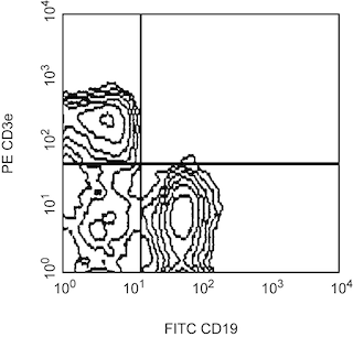
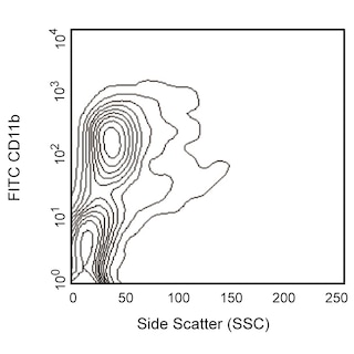
The 6C1 monoclonal antibody specifically recognizes the common epitopes of PIR-A and PIR-B (Paired Immunoglobulin-liked Receptors) in all mouse strains tested (A/J, BALB/cJ, C3H, C57BL/6, DBA/1, DBA/2, NZB, and SJL). PIR-A and PIR-B are type-I transmembrane glycoproteins containing six Ig-like domains. There are multiple PIR-A proteins which are activating receptors by virtue of their intracellular association with the ITAM (Immunoreceptor Tyrosine-based Activation Motif)- containing Fc receptor γ chain (FcRγ) in mast cells and macrophages. FcRγ expression is required for PIR-A cell-surface expression on dendritic cells, B cells, and myeloid lineages. In contrast, PIR-B is an inhibitory receptor which contains various ITIMs (Immunoreceptor Tyrosine-based Inhibitory Motifs) in its cytoplasmic domain. These receptors are expressed on B cells, granulocytes, mast cells, dendritic cells, and monocytes/macrophages, but not on thymocytes, T lymphocytes, erythroid lineage cells, or NK cells. The level of cell-surface expression of these receptors increases as a function of B-cell activation and myeloid- and B-lineage differentiation. The 6C1 antibody immunoprecipitates molecules of 85- and 125-kDa, which correspond to PIRA and PIR-B, respectively.

Development References (6)
-
Bléry M, Kubagawa H, Chen CC, Vély F, Cooper MD, Vivier E. The paired Ig-like receptor PIR-B is an inhibitory receptor that recruits the protein-tyrosine phosphatase SHP-1. Proc Natl Acad Sci U S A. 1998; 95(5):2446-2451. (Biology). View Reference
-
Chen CC, Hurez V, Brockenbrough JS, Kubagawa H, Cooper MD. Paternal monoallelic expression of the paired immunoglobulin-like receptors PIR-A and PIR-B. Proc Natl Acad Sci U S A. 1999; 96(12):6868-6872. (Clone-specific). View Reference
-
Kubagawa H, Burrows PD, Cooper MD. A novel pair of immunoglobulin-like receptors expressed by B cells and myeloid cells. Proc Natl Acad Sci U S A. 1997; 94(10):5261-5266. (Biology). View Reference
-
Kubagawa H, Chen CC, Ho LH. Biochemical nature and cellular distribution of the paired immunoglobulin-like receptors, PIR-A and PIR-B. J Exp Med. 1999; 189(2):309-318. (Immunogen). View Reference
-
Maeda A, Kurosaki M, Kurosaki T. Paired immunoglobulin-like receptor (PIR)-A is involved in activating mast cells through its association with Fc receptor gamma chain. J Exp Med. 1998; 188(5):991-995. (Biology). View Reference
-
Taylor LS, McVicar DW. Functional association of FcepsilonRIgamma with arginine(632) of paired immunoglobulin-like receptor (PIR)-A3 in murine macrophages. Blood. 1999; 94(5):1790-1796. (Biology). View Reference
Please refer to Support Documents for Quality Certificates
Global - Refer to manufacturer's instructions for use and related User Manuals and Technical data sheets before using this products as described
Comparisons, where applicable, are made against older BD Technology, manual methods or are general performance claims. Comparisons are not made against non-BD technologies, unless otherwise noted.
For Research Use Only. Not for use in diagnostic or therapeutic procedures.