Old Browser
This page has been recently translated and is available in French now.
Looks like you're visiting us from {countryName}.
Would you like to stay on the current country site or be switched to your country?


.png)

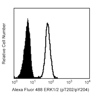
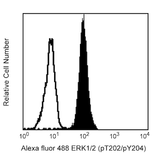
Analysis of Syk (pY348) in human peripheral blood lymphocytes. Human peripheral blood mononuclear cells (PBMC) were either stimulated (right panel) by cross-linking of surface IgM with purified Goat F(ab')2 Anti-Human IgM (SouthernBiotech) at 37°C for 8 minutes or unstimulated (left panel). The cells were fixed with pre-warmed BD Phosflow™ Lyse/Fix Buffer (Cat. No. 558049) for 10 minutes at 37ºC, permeabilized with BD Phosflow™ Perm Buffer II (Cat. No. 558052) at room temperature for 10 minutes, and then stained with PE Mouse anti-Syk (pY348) and Alexa Fluor® 647 Mouse anti-human CD20 mAb H1(FB1) (Cat. No. 558054). For data analysis, lymphocytes were selected by their scatter profile. The data demonstrates that the upregulated phosphorylation is restricted to the stimulated CD20-positive B lymphocytes. Flow cytometry was performed on a BD FACSCalibur™ flow cytometry system.

.png)

BD™ Phosflow PE Mouse anti-Syk (pY348)

BD™ Phosflow PE Mouse anti-Syk (pY348)
.png)
Regulatory Status Legend
Any use of products other than the permitted use without the express written authorization of Becton, Dickinson and Company is strictly prohibited.
Preparation And Storage
Recommended Assay Procedures
This antibody conjugate is suitable for intracellular staining of human lymphoid cell lines, peripheral blood mononuclear cells, and mouse splenocytes using BD Phosflow™ Fix Buffer I or Lyse/Fix Buffer. Any of the three BD Phosflow™ permeabilization buffers may be used.
Product Notices
- This reagent has been pre-diluted for use at the recommended Volume per Test. We typically use 1 × 10^6 cells in a 100-µl experimental sample (a test).
- Please refer to www.bdbiosciences.com/us/s/resources for technical protocols.
- For fluorochrome spectra and suitable instrument settings, please refer to our Multicolor Flow Cytometry web page at www.bdbiosciences.com/colors.
- Caution: Sodium azide yields highly toxic hydrazoic acid under acidic conditions. Dilute azide compounds in running water before discarding to avoid accumulation of potentially explosive deposits in plumbing.
- Source of all serum proteins is from USDA inspected abattoirs located in the United States.
Companion Products


Syk is a non-receptor protein-tyrosine kinase that is closely related to ZAP70 and plays crucial roles in the development and receptor-mediated signaling of most leukocytes and in vascular integrity. Syk is expressed in hematopoietic cells, including B lymphocytes, immature (CD4, CD8 double-negative and double-positive) thymocytes, and myeloid cells, epithelial cell lines, and normal breast tissue. Mature (CD4 or CD8 single-positive) thymocytes and peripheral αβ TCR-bearing T lymphocytes have very low or undetectable levels of Syk. Syk contributes to the signal transduction process by binding to ITAMs (Immunoreceptor Tyrosine-based Activation Motifs) of immune receptors, including Igα and Igβ (CD79a and b), TCRζ, CD3ε, and FcRγ. Upon receptor activation, Syk binds to phosphorylated ITAMs via its two N-terminal SH2 domains thereby activating Syk and causing tyrosines in the interdomain, between the SH2 and Kinase domains of Syk, to undergo auto-phosphorylation and phosphorylation by Lyn. The tyrosine 348 phosphorylation site (pY348) in human Syk is orthologous to tyrosine 342 in mouse and rat Syk and tyrosine 315 in human ZAP70. This phosphorylated site can act as a binding site for other signaling molecules, such as PLCγ, Vav, and Fgr.
The I120-772 antibody is specific for human Syk (pY348) and does not cross-react with phosphorylated Zap70. The orthologous phosphorylation site in mouse and rat Syk is Y342.

Development References (7)
-
Abtahian F, Guerriero A, Sebzda E, et al. . Regulation of blood and lymphatic vascular separation by signaling proteins SLP-76 and Syk. Science. 2003; 299:247-251. (Biology).
-
Coopman PJP, Do MTH, Barth M, et al. The Syk tyrosine kinase suppresses malignant growth of human breast cancer cells. Nature. 2000; 406:742-747. (Biology).
-
Hong JJ, Yankee TM, Harrison ML, Geahlen RL. Regulation of signaling in B cells through the phosphorylation of Syk on linker region tyrosines. J Biol Chem. 2002; 277:31703-31714. (Biology).
-
Latour S, Veillette A. Proximal protein tyrosine kinases in immunoreceptor signaling. Curr Opin Immunol. 2001; 13:299-306. (Biology).
-
Turner M, Schweighoffer E, Colucci F, Di Santo JP, Tybulewicz VL. Tyrosine kinase SYK; essential functions for immunoreceptor signaling. Immunol Today. 2000; 21:148-154. (Biology). View Reference
-
Vines CM, Potter JW, Xu Y, et al. Inhibition of β2 integrin receptor and Syk kinase signaling in monocytes by the Src family kinase Fgr. Immunity. 2001; 15:507-519. (Biology).
-
Zhang J, Benestein E, Siraganian RP. Phosphorylation of Tyr342 in the linker region of Syk is critical for FceRI signaling in mast cells. Mol Cell Biol. 2002; 22:8144-8154. (Biology).
Please refer to Support Documents for Quality Certificates
Global - Refer to manufacturer's instructions for use and related User Manuals and Technical data sheets before using this products as described
Comparisons, where applicable, are made against older BD Technology, manual methods or are general performance claims. Comparisons are not made against non-BD technologies, unless otherwise noted.
For Research Use Only. Not for use in diagnostic or therapeutic procedures.