Old Browser
This page has been recently translated and is available in French now.
Looks like you're visiting us from {countryName}.
Would you like to stay on the current country site or be switched to your country?


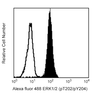
Analysis of S6 (pS244) in activated human peripheral blood mononuclear cells (PBMC). PBMC were isolated by density gradient centrifugation (Ficoll-Paque™ PLUS, Cat. No. 17-1440-02) and either left untreated (open histogram) or treated with PMA (Sigma-Aldrich, Cat. No. P8139) at 50 nM/10^6 cells for 30 minutes (shaded histogram). Cells were then fixed in BD Cytofix™ buffer (Cat. No. 554655) at 37°C for 10 minutes, then permeabilized with BD Phosflow™ Perm Buffer III (Cat. No. 558050) on ice for at least 30 minutes, and then stained with Alexa Fluor® 647 Mouse anti-S6 (pS244, Cat. No. 560645). For data analysis, lymphocytes were selected by scatter profile. Flow cytometry was performed on a BD FACSCalibur™ flow cytometry system.


Western blot analysis of S6 (pS244). The specificity of mAb N5-676 was confirmed by western blot analysis using unconjugated Mouse anti-S6 (pS244) antibody on lysates from untreated (lane 1) or PMA-treated (lane 2) PBMC. S6 (pS244) is identified as a band of 32 kDa, with increased intensity in the PMA-treated cells. Quantitative western blot analyses demonstrated that the unidentified high-molecular-weight bands are not induced by the PMA treatment (data not shown) and, thus, do not contribute to the signal increase that is observed by flow cytometry. Purified Mouse anti-Actin monoclonal antibody (Cat. No. 612656/612657) was the gel-loading control.


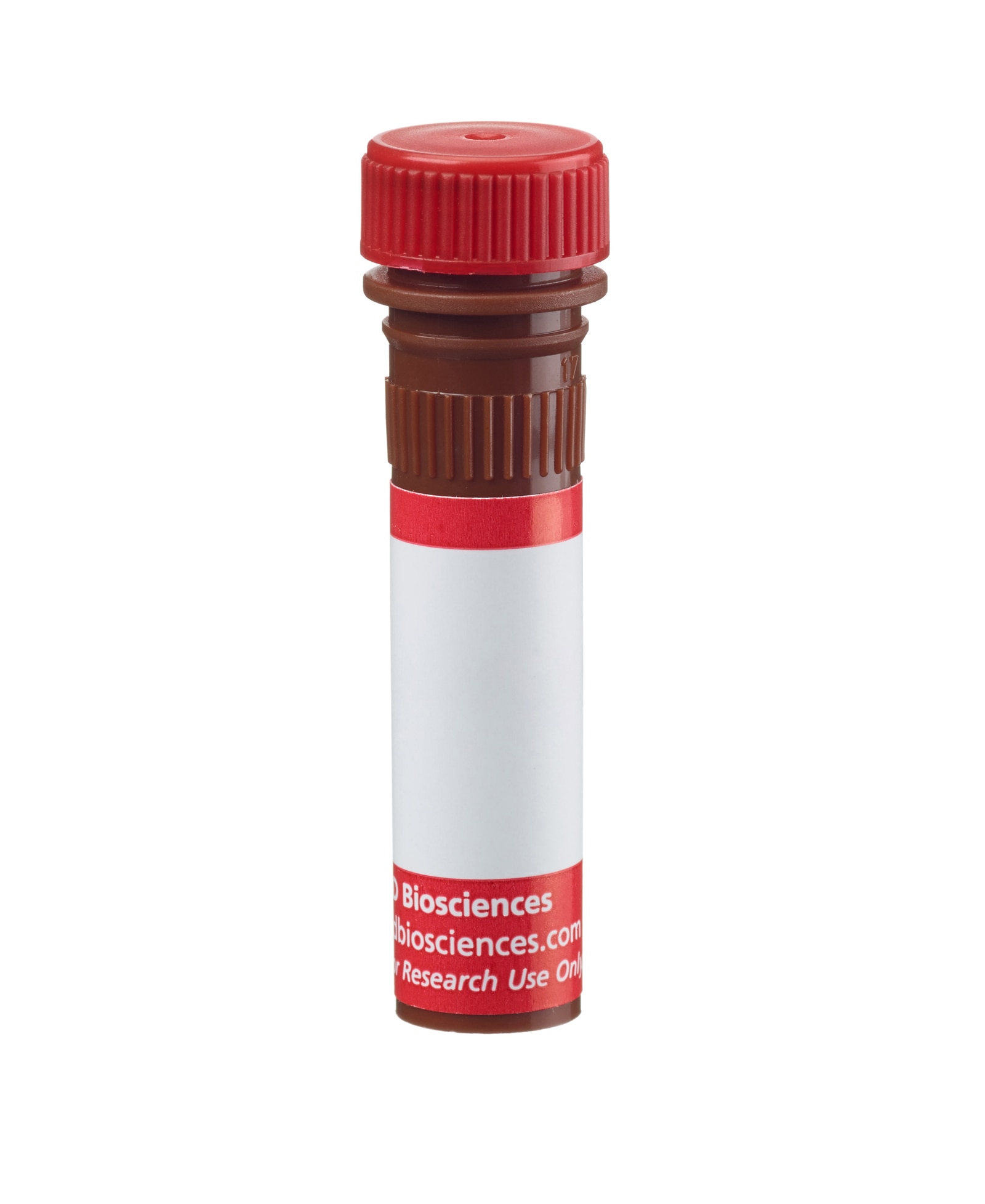
BD™ Phosflow Alexa Fluor® 647 Mouse anti-S6 (pS244)

BD™ Phosflow Alexa Fluor® 647 Mouse anti-S6 (pS244)

BD™ Phosflow Alexa Fluor® 647 Mouse anti-S6 (pS244)

Regulatory Status Legend
Any use of products other than the permitted use without the express written authorization of Becton, Dickinson and Company is strictly prohibited.
Preparation And Storage
Recommended Assay Procedures
This antibody conjugate is suitable for intracellular staining of human peripheral blood mononuclear cells using BD Cytofix™ Fixation Buffer. Any of the three BD Phosflow™ permeabilization buffers may be used.
Product Notices
- This reagent has been pre-diluted for use at the recommended Volume per Test. We typically use 1 × 10^6 cells in a 100-µl experimental sample (a test).
- Source of all serum proteins is from USDA inspected abattoirs located in the United States.
- Caution: Sodium azide yields highly toxic hydrazoic acid under acidic conditions. Dilute azide compounds in running water before discarding to avoid accumulation of potentially explosive deposits in plumbing.
- Alexa Fluor® 647 fluorochrome emission is collected at the same instrument settings as for allophycocyanin (APC).
- For fluorochrome spectra and suitable instrument settings, please refer to our Multicolor Flow Cytometry web page at www.bdbiosciences.com/colors.
- The Alexa Fluor®, Pacific Blue™, and Cascade Blue® dye antibody conjugates in this product are sold under license from Molecular Probes, Inc. for research use only, excluding use in combination with microarrays, or as analyte specific reagents. The Alexa Fluor® dyes (except for Alexa Fluor® 430), Pacific Blue™ dye, and Cascade Blue® dye are covered by pending and issued patents.
- Ficoll-Paque is a trademark of Amersham Biosciences Limited.
- Alexa Fluor® is a registered trademark of Molecular Probes, Inc., Eugene, OR.
- Please refer to www.bdbiosciences.com/us/s/resources for technical protocols.
Companion Products


Ribosomal protein S6 (~29 kDa calculated and ~32 kDa observed molecular weights) is a component of the 40S ribosomal subunit and belongs to the S6E family of ribosomal proteins. The S6 ribosomal protein plays a role in regulating the translation of RNAs and thus controlling the growth and proliferation of cells. S6 ribosomal protein phosphorylation, especially at multiple C-terminal serine residues S235, S236, S240, and S244, activates S6. The activated S6 ribosomal protein in turn upregulates the ribosomal translation of RNA species coding for other ribosomal proteins, peptide elongation factors and other proteins involved in cell cycle entry and progression. These phosphorylations are mediated by various kinases (e.g., p70S6K and PKCD) activated through cellular responses to growth factors, cytokines, tumor promoting agents, and mitogens. The S6 ribosomal protein can be dephosphorylated in growth-arrested cells.
The N5-676 monoclonal antibody specifically detects the S6 ribosomal protein phosphorylated at S244.
Development References (7)
-
Corradetti MN, Inoki K, Guan KL. The stress-inducted proteins RTP801 and RTP801L are negative regulators of the mammalian target of rapamycin pathway. J Biol Chem. 2005; 280(11):9769-9772. (Biology). View Reference
-
Gu JJ, Santiago L, Mitchell BS. Synergy between imatinib and mycophenolic acid in inducing apoptosis in cell lines expressing Bcr-Abl. Blood. 2005; 105(8):3270-3277. (Biology). View Reference
-
Jastrzebski K, Hannan KM, Tchoubrieva EB, Hannan RD, Pearson RB. Coordinate regulation of ribosome biogenesis and function by the ribosomal protein S6 kinase, a key mediator of mTOR function. Growth Factors. 2007; 25(4):209-226. (Biology). View Reference
-
Kleijn M, Proud CG. The regulation of protein synthesis and translation factors by CD3 and CD28 in human primary T lymphocytes. BMC Biochem. 2002; 3:11. (Biology). View Reference
-
Lal L, Li Y, Smith J, et al. Activation of the p70 S6 kinase by all-trans-retinoic acid in acute promyelocytic leukemia cells.. Blood. 2005; 105(4):1669-77. (Biology). View Reference
-
Shah OJ, Ghosh S, Hunter T. Mitotic regulation of ribosomal S6 kinase 1 involves Ser/Thr, Pro phosphorylation of consensus and non-consensus sites by Cdc2. J Biol Chem. 2003; 278(18):16433-16442. (Biology). View Reference
-
Shah OJ, Hunter T. Critical role of T-loop and H-motif phosphorylation in the regulation of S6 kinase 1 by the tuberous sclerosis complex. J Biol Chem. 2004; 279(20):20816-20823. (Biology). View Reference
Please refer to Support Documents for Quality Certificates
Global - Refer to manufacturer's instructions for use and related User Manuals and Technical data sheets before using this products as described
Comparisons, where applicable, are made against older BD Technology, manual methods or are general performance claims. Comparisons are not made against non-BD technologies, unless otherwise noted.
For Research Use Only. Not for use in diagnostic or therapeutic procedures.
Report a Site Issue
This form is intended to help us improve our website experience. For other support, please visit our Contact Us page.