Dear Customer, If you experience any text readability or display issues on our website, please update your web browser to the latest version
.
See More
Dear Customer, If you experience any text readability or display issues on our website, please update your web browser to the latest version
.
See MoreDear Customer, If you experience any text readability or display issues on our website, please update your web browser to the latest version
.
This page has been recently translated and is available in French now.
Looks like you're visiting us from {countryName}.
Would you like to stay on the current location site or be switched to your location?
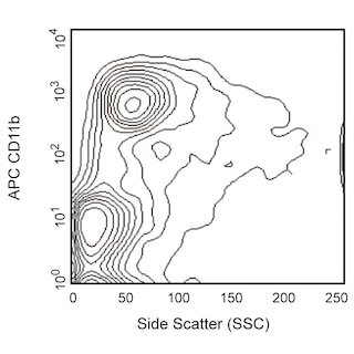
Immunohistofluorescent analysis of I-A/I-E expression by cells within C57BL/6 mouse spleen (Left Panel). A mouse spleen cryosection (5 µm) was fixed with BD Cytofix™ Fixation Buffer (Cat. No. 554655), blocked with 5% goat serum and 1% BSA diluted in 1x PBS, and stained with Alexa Fluor® 488 Rat Anti-Mouse I-A/I-E antibody (Cat. No. 562352, pseudo-colored green) and Alexa Fluor® 647 Rat Anti-Mouse CD3 Molecular Complex antibody (Cat. No. 557869, pseudo-colored blue). Images were captured on a standard epifluorescence microscope. Original magnification, 20x. Mouse spleen cells from either M5/114-negative SJL (Middle Panel) or M5/114-positive BALB/c (Right Panel) mice were stained with Alexa Fluor® 488 Rat Anti-Mouse I-A/I-E (Cat. No. 562352), APC Rat Anti-Mouse CD45R/B220 (Cat. No. 553092/561880) and APC Rat Anti-Mouse CD11b (Cat. No. 553312/561690) antibodies. Two-color flow cytometric dot plots showing the expression of I-A/I-E MHC class II alloantigens versus CD45R/B220 and CD11b were derived from gated events with the forward and side light-scatter characteristics of viable splenocytes. The M5/114 monoclonal antibody detects I-Ad and I-Ed MHC class II alloantigens that are expressed on both B cells and macrophages. Flow cytometry was performed using a BD™ LSR II Flow Cytometer System.
Any use of products other than the permitted use without the express written authorization of Becton, Dickinson and Company is strictly prohibited.
The M5/114.15.2 monoclonal antibody recognizes a polymorphic determinant shared by the I-A[b], I-A[d], I-A[q], I-E[d], and I-E[k] (but not I-A[f], I-A[k], or I-A[s]) MHC class II alloantigens that can be expressed by B cells, dendritic cells, monocytes, macrophages and activated T cells. It also reacts with cells from mice of the H-2[p] and H-2[r] haplotypes, and it is non-reactive with cells from NOD (H-2[g7]) mice. Flow cytometric analysis indicates that the M5/114.15.2 and 2G9 monoclonal antibodies have comparable reactivity on cells from mice with I-A[b], I-A[d], I-A[g7], I-A[q], I-E[d], and I-E[k] alloantigens.
Please refer to Support Documents for Quality Certificates
Global - Refer to manufacturer's instructions for use and related User Manuals and Technical data sheets before using this products as described
Comparisons, where applicable, are made against older BD Technology, manual methods or are general performance claims. Comparisons are not made against non-BD technologies, unless otherwise noted.
For Research Use Only. Not for use in diagnostic or therapeutic procedures.