-
Reagents
- Flow Cytometry Reagents
-
Western Blotting and Molecular Reagents
- Immunoassay Reagents
-
Single-Cell Multiomics Reagents
- BD® OMICS-Guard Sample Preservation Buffer
- BD® AbSeq Assay
- BD® Single-Cell Multiplexing Kit
- BD Rhapsody™ ATAC-Seq Assays
- BD Rhapsody™ Whole Transcriptome Analysis (WTA) Amplification Kit
- BD Rhapsody™ TCR/BCR Next Multiomic Assays
- BD Rhapsody™ Targeted mRNA Kits
- BD Rhapsody™ Accessory Kits
- BD® OMICS-One Protein Panels
-
Functional Assays
-
Microscopy and Imaging Reagents
-
Cell Preparation and Separation Reagents
-
- BD® OMICS-Guard Sample Preservation Buffer
- BD® AbSeq Assay
- BD® Single-Cell Multiplexing Kit
- BD Rhapsody™ ATAC-Seq Assays
- BD Rhapsody™ Whole Transcriptome Analysis (WTA) Amplification Kit
- BD Rhapsody™ TCR/BCR Next Multiomic Assays
- BD Rhapsody™ Targeted mRNA Kits
- BD Rhapsody™ Accessory Kits
- BD® OMICS-One Protein Panels
- France (English)
-
Change country/language
Old Browser
This page has been recently translated and is available in French now.
Looks like you're visiting us from United States.
Would you like to stay on the current country site or be switched to your country?
BD Cytoperm™ Permeabilization Buffer Plus

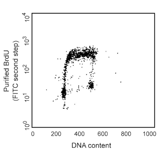
Flow cytometric analysis of DNA synthesis by TK-1 cells. TK-1 cells were either pulsed with 50 µM BrdU for 1 hour (left panel) or were not pulsed (right panel). Staining was performed using BD Cytoperm™ Permeabilization Buffer Plus in the procedure from the BD Pharmingen™ FITC and APC BrdU Flow Kits. The permeabilized cells were stained with the PerCP-Cy™5.5 Mouse Anti-BrdU monoclonal antibody (Cat. No. 560809) followed by the DNA-specific dye, DAPI dihydrochloride at 1 µg/mL (Sigma, Cat. No. D9542). Two-color flow cytometric dot plots showing the correlated expression patterns of DAPI vs BrdU were derived from gated events with the forward and side light-scatter characteristics of viable lymphocytes. Flow cytometry was performed with doublet discrimination using a BD™ LSRII system.

Flow cytometric analysis of DNA synthesis by TK-1 cells. TK-1 cells were either pulsed with 50 µM BrdU for 1 hour (left panel) or were not pulsed (right panel). Staining was performed using BD Cytoperm™ Permeabilization Buffer Plus in the procedure from the BD Pharmingen™ FITC and APC BrdU Flow Kits. The permeabilized cells were stained with the PerCP-Cy™5.5 Mouse Anti-BrdU monoclonal antibody (Cat. No. 560809) followed by the DNA-specific dye, DAPI dihydrochloride at 1 µg/mL (Sigma, Cat. No. D9542). Two-color flow cytometric dot plots showing the correlated expression patterns of DAPI vs BrdU were derived from gated events with the forward and side light-scatter characteristics of viable lymphocytes. Flow cytometry was performed with doublet discrimination using a BD™ LSRII system.

Flow cytometric analysis of DNA synthesis by TK-1 cells. TK-1 cells were either pulsed with 50 µM BrdU for 1 hour (left panel) or were not pulsed (right panel). Staining was performed using BD Cytoperm™ Permeabilization Buffer Plus in the procedure from the BD Pharmingen™ FITC and APC BrdU Flow Kits. The permeabilized cells were stained with the PerCP-Cy™5.5 Mouse Anti-BrdU monoclonal antibody (Cat. No. 560809) followed by the DNA-specific dye, DAPI dihydrochloride at 1 µg/mL (Sigma, Cat. No. D9542). Two-color flow cytometric dot plots showing the correlated expression patterns of DAPI vs BrdU were derived from gated events with the forward and side light-scatter characteristics of viable lymphocytes. Flow cytometry was performed with doublet discrimination using a BD™ LSRII system.

BD Cytofix/Cytoperm™ Plus Permeabilization Buffer Plus
Regulatory Status Legend
Any use of products other than the permitted use without the express written authorization of Becton, Dickinson and Company is strictly prohibited.
Product Details
Description
BD Cytoperm™ Permeabilization Buffer Plus is specially formulated for the immunofluorescent staining of incorporated BrdU for flow cytometric analysis. It is used as a staining enhancer and secondary permeabilization reagent. BD Cytoperm™ Permeabilization Buffer Plus should be used with fixed cell samples only. Use of this buffer on unfixed cells will cause cell damage.
Preparation And Storage
Recommended Assay Procedures
BD Cytoperm™ Permeabilization Buffer Plus is specially formulated for the immunofluorescent staining of incorporated BrdU for flow cytometric analysis and may be found in the BD Pharmingen™ FITC BrdU Flow Kit (Cat. No. 559619 / 557891) or the BD Pharmingen™ APC BrdU Flow Kit (Cat. No. 552598 / 557892). Investigators may find the following abbreviated protocol to be helpful.
1. Immunofluorescent staining of cell surface antigens.
a. Add BrdU-pulsed cells (10^6 cells in 50 µL of staining buffer) to flow cytometry tubes.
b. Add fluorescent antibodies specific for cell-surface markers in 50 µL of staining buffer (eg, BD Pharmingen™ Stain Buffer (FBS) Cat. No. 554656) per tube and mix well.
c. Incubate cells with antibodies for 15 minutes on ice.
d. Wash cells 1x by adding 1 mL of staining buffer per tube, centrifuge (5 min.) at 200 - 300 x g, and discard supernatant.
2. Fix and permeabilize cells with BD Cytofix/Cytoperm Buffer.
a. Resuspend cells with 100 µL of BD Cytofix/Cytoperm Buffer per tube.
b. Incubate cells for 15 - 30 minutes at room temperature or on ice.
c. Wash cells 1x with 1 mL of 1x BD Perm/Wash Buffer, centrifuge as in step 1d and discard supernatant.
3. Incubate cells with BD Cytoperm™ Permeabilization Buffer Plus.
a. Resuspend cells with 100 µL of BD Cytoperm™ Permeabilization Buffer Plus per tube.
b. Incubate cells for 10 minutes on ice.
c. Wash cells 1x by adding 1 mL of 1x BD Perm/Wash Buffer (as in Step 2c).
4. Re-Fixation of cells
a. Resuspend cells with 100 µL of BD Cytofix/Cytoperm Buffer per tube.
b. Incubate cells for 5 minutes at room temperature or on ice.
c. Wash cells 1x by adding 1 mL of 1x BD Perm/Wash Buffer (as in Step 2c).
5. Treatment of cells with DNase to expose incorporated BrdU.
a. Resuspend cells with 100 µL of diluted DNase (diluted to 300 µg/mL in DPBS) per tube, (ie, 30 µg of DNase to each tube).
b. Incubate cells for 1 hour at 37°C.
c. Wash cells 1x by adding 1 mL of 1x BD Perm/Wash Buffer (as in Step 2c).
6. Stain BrdU and intracellular antigens with fluorescent antibodies.
a. Resuspend cells with 50 µL of BD Perm/Wash Buffer containing diluted fluorescent anti-BrdU and/or antibodies specific for intracellular antigens.
b. Incubate cells for 20 minutes at room temperature.
c. Wash cells 1x by adding 1 mL of 1x BD Perm/Wash Buffer (as in Step 2c).
7. Optional - Staining of total DNA for cell cycle analysis.
Note: Proceed to Step 8 if the staining of total DNA levels is not desired.
a. Resuspend cells with 20 µL of the 7-AAD solution (Cat. No. 559925).
8. Resuspension of cells for Flow Cytometric Analysis.
a. Add 1 mL of staining buffer to each tube to resuspend cells.
b. Analyze stained cells with a flow cytometer (run at a rate no greater than 400 events/sec.) and acquire multiparameter data files.
Note: Samples may be stored overnight at 4°C, protected from exposure to light, prior to analysis by flow cytometry.
Product Notices
- Source of all serum proteins is from USDA inspected abattoirs located in the United States.
- Caution: Sodium azide yields highly toxic hydrazoic acid under acidic conditions. Dilute azide compounds in running water before discarding to avoid accumulation of potentially explosive deposits in plumbing.
- Cy is a trademark of GE Healthcare.
- Please refer to www.bdbiosciences.com/us/s/resources for technical protocols.
Data Sheets
Companion Products


Development References (1)
-
BD Pharmingen™. BrdU Flow Kits Instruction Manual. San Jose, CA: BD Biosciences; 2008:1-40.
Please refer to Support Documents for Quality Certificates
Global - Refer to manufacturer's instructions for use and related User Manuals and Technical data sheets before using this products as described
Comparisons, where applicable, are made against older BD Technology, manual methods or are general performance claims. Comparisons are not made against non-BD technologies, unless otherwise noted.
For Research Use Only. Not for use in diagnostic or therapeutic procedures.