How Dyes Evolved: The History of the Modern Flow Cytometry Dye
September 12, 2022
Dyes are an important part of flow cytometry. The sophisticated, multi-colour flow cytometry experiments being done today are not possible without state-of-the-art, high-performance dyes. The evolution from the early fluorescence dyes to the highly sophisticated polymer dyes of today that have enabled advanced immunophenotyping experiments, is a very exciting story. Advances in instrumentation and laser technology are also important aspects that have brought about these developments. Read about this story and learn how chemistry, engineering and science have been working together over the years to make modern flow cytometry possible.
The Flow Cytometry Dye Timeline
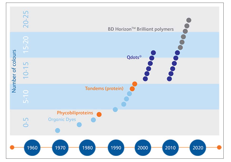
How the dye, a vital component of flow cytometry, has evolved over the ages1
| Early 1970s | Only two fluorescent dyes were available – Fluorescein and rhodamine. |
| Early 1980s | Phycobiliproteins like phycoerythrin (PE) and allophycocyanin (APC), extracted from cyanobacteria and algae, were developed by Vernon Oi, Alex Glazer and Lubert Stryer. |
| Late 1980s | The ability of PE to absorb and transfer energy to other fluorescent molecules was used to create tandem dyes (eg: PE-Texas Red, PE-Cy5, PE-Cy5.5, PE-Cy7). |
| 1990s | APC-based tandem dyes synthesized. Alexa™ dyes, which are a large spectrally-resolved series of small organic dyes, became available. 8 - 11 colour polychromatic flow cytometry became possible. |
| Early 2000s | Introduction of fluorescent, semiconductor nanocrystals (quantum dots or Qdots™) enabled 18-colour flow-cytometry. |
| 2011 and later | BD Horizon™ Brilliant violet and ultraviolet dyes have become available, which are developed using organic polymers. These dyes and their tandems provide additional bright options at a variety of wave¬lengths and are more suitable for immunophenotyping than quantum dots overcoming the main issues Qdots™ were causing. |