BD Pharmingen™ Purified Mouse Anti-Human Cdc25C
Clone C2-2 (RUO)
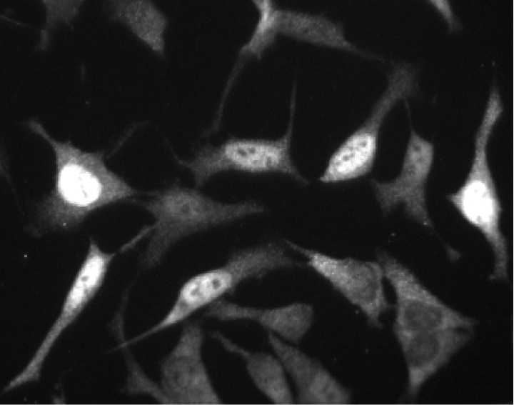
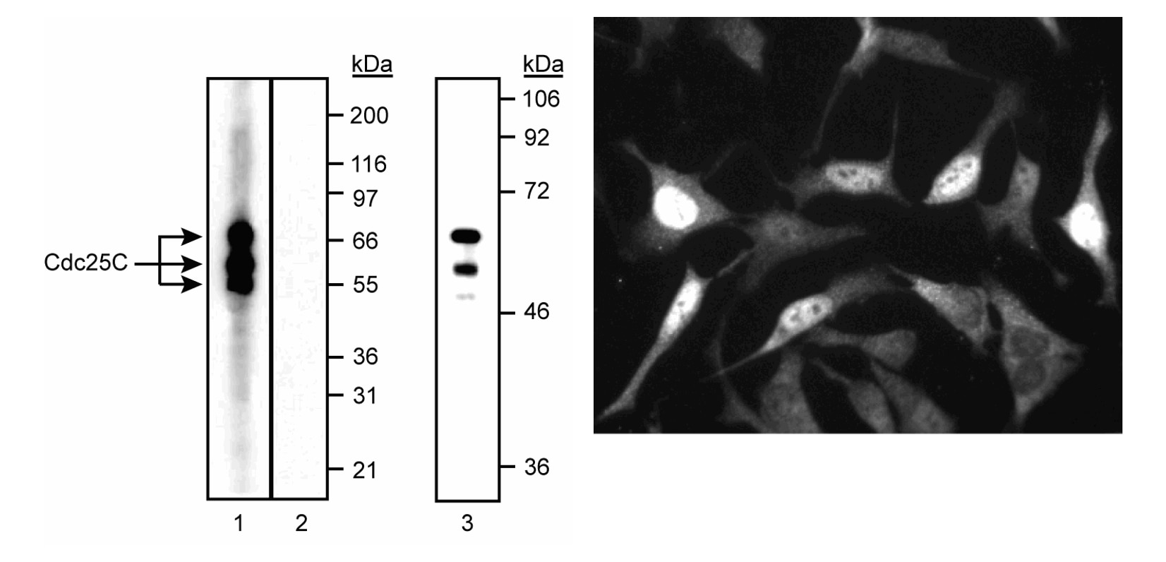
Left: Western blot analysis of Cdc25C in human and mouse cell lysates. Lanes 1 and 2, A549 human lung carcinoma cells were probed with clone C2-2 at 1-2 µg/ml (lane 1) or with an isotype control (lane 2). Lane 3, Rous Sarcoma Virsu-transformed mouse 3T3 cells. The C2-2 antibody identifies Cdc25C isoforms ranging between 60-63 kDa in both human and mouse cell types. Right: Immunofluorescent staining of A549 (ATCC CCL-185) cells. Cells were seeded in a 96 well imaging plate (Ca. No. 353219) at ~ 10 000 cells per well. After overnight incubation, cells were stained using the Triton™ X-100 perm protocol and the anti-Cdc25C antibody. The second step reagent was FITC goat anti mouse Ig (Cat. No. 554001). Images were taken on a BD Pathway™ 855 bioimaging system using a 20x objective. This antibody also stained HeLa (ATCC CCL-2) and U-2 OS (ATCC HTB-96) cells with either the Triton™ X-100 or alcohol perm protocols (see Recommended Assay Procedure).
Left: Western blot analysis of Cdc25C in human and mouse cell lysates. Lanes 1 and 2, A549 human lung carcinoma cells were probed with clone C2-2 at 1-2 µg/ml (lane 1) or with an isotype control (lane 2). Lane 3, Rous Sarcoma Virsu-transformed mouse 3T3 cells. The C2-2 antibody identifies Cdc25C isoforms ranging between 60-63 kDa in both human and mouse cell types. Right: Immunofluorescent staining of A549 (ATCC CCL-185) cells. Cells were seeded in a 96 well imaging plate (Ca. No. 353219) at ~ 10 000 cells per well. After overnight incubation, cells were stained using the Triton™ X-100 perm protocol and the anti-Cdc25C antibody. The second step reagent was FITC goat anti mouse Ig (Cat. No. 554001). Images were taken on a BD Pathway™ 855 bioimaging system using a 20x objective. This antibody also stained HeLa (ATCC CCL-2) and U-2 OS (ATCC HTB-96) cells with either the Triton™ X-100 or alcohol perm protocols (see Recommended Assay Procedure).

Left: Western blot analysis of Cdc25C in human and mouse cell lysates. Lanes 1 and 2, A549 human lung carcinoma cells were probed with clone C2-2 at 1-2 µg/ml (lane 1) or with an isotype control (lane 2). Lane 3, Rous Sarcoma Virsu-transformed mouse 3T3 cells. The C2-2 antibody identifies Cdc25C isoforms ranging between 60-63 kDa in both human and mouse cell types. Right: Immunofluorescent staining of A549 (ATCC CCL-185) cells. Cells were seeded in a 96 well imaging plate (Ca. No. 353219) at ~ 10 000 cells per well. After overnight incubation, cells were stained using the Triton™ X-100 perm protocol and the anti-Cdc25C antibody. The second step reagent was FITC goat anti mouse Ig (Cat. No. 554001). Images were taken on a BD Pathway™ 855 bioimaging system using a 20x objective. This antibody also stained HeLa (ATCC CCL-2) and U-2 OS (ATCC HTB-96) cells with either the Triton™ X-100 or alcohol perm protocols (see Recommended Assay Procedure).
Left: Western blot analysis of Cdc25C in human and mouse cell lysates. Lanes 1 and 2, A549 human lung carcinoma cells were probed with clone C2-2 at 1-2 µg/ml (lane 1) or with an isotype control (lane 2). Lane 3, Rous Sarcoma Virsu-transformed mouse 3T3 cells. The C2-2 antibody identifies Cdc25C isoforms ranging between 60-63 kDa in both human and mouse cell types. Right: Immunofluorescent staining of A549 (ATCC CCL-185) cells. Cells were seeded in a 96 well imaging plate (Ca. No. 353219) at ~ 10 000 cells per well. After overnight incubation, cells were stained using the Triton™ X-100 perm protocol and the anti-Cdc25C antibody. The second step reagent was FITC goat anti mouse Ig (Cat. No. 554001). Images were taken on a BD Pathway™ 855 bioimaging system using a 20x objective. This antibody also stained HeLa (ATCC CCL-2) and U-2 OS (ATCC HTB-96) cells with either the Triton™ X-100 or alcohol perm protocols (see Recommended Assay Procedure).
Preparation And Storage
Recommended Assay Procedures
Bioimaging
1. Seed the cells in appropriate culture medium at ~10,000 cells per well in a BD Falcon™ 96-well Imaging Plate (Cat. No. 353219) and culture overnight.
2. Remove the culture medium from the wells, and fix the cells by adding 100 μl of BD Cytofix™ Fixation Buffer (Cat. No. 554655) to each well. Incubate for 10 minutes at room temperature (RT).
3. Remove the fixative from the wells, and permeabilize the cells using either BD Perm Buffer III, 90% methanol, or Triton™ X-100:
a. Add 100 μl of -20°C 90% methanol or Perm Buffer III (Cat. No. 558050) to each well and incubate for 5 minutes at RT.
OR
b. Add 100 μl of 0.1% Triton™ X-100 to each well and incubate for 5 minutes at RT.
4. Remove the permeabilization buffer, and wash the wells twice with 100 μl of 1× PBS.
5. Remove the PBS, and block the cells by adding 100 μl of BD Pharmingen™ Stain Buffer (FBS) (Cat. No. 554656) to each well. Incubate for 30 minutes at RT.
6. Remove the blocking buffer and add 50 μl of the optimally titrated primary antibody (diluted in Stain Buffer) to each well, and incubate for 1 hour at RT.
7. Remove the primary antibody, and wash the wells three times with 100 μl of 1× PBS.
8. Remove the PBS, and add the second step reagent at its optimally titrated concentration in 50 μl to each well, and incubate in the dark for 1 hour at RT.
9. Remove the second step reagent, and wash the wells three times with 100 μl of 1× PBS.
10. Remove the PBS, and counter-stain the nuclei by adding 200 μl per well of 2 μg/ml Hoechst 33342 (e.g., Sigma-Aldrich Cat. No. B2261) in 1× PBS to each well at least 15 minutes before imaging.
11. View and analyze the cells on an appropriate imaging instrument.
Bioimaging: For more detailed information please refer to http://www.bdbiosciences.com/support/resources/protocols/ceritifed_reagents.jsp
Western blot: For more detailed information please refer to http://www.bdbiosciences.com/support/resources/protocols/monoclonal_anti.jsp
Product Notices
- Since applications vary, each investigator should titrate the reagent to obtain optimal results.
- This antibody has been developed and certified for the bioimaging application. However, a routine bioimaging test is not performed on every lot. Researchers are encouraged to titrate the reagent for optimal performance.
- Triton is a trademark of the Dow Chemical Company.
- Caution: Sodium azide yields highly toxic hydrazoic acid under acidic conditions. Dilute azide compounds in running water before discarding to avoid accumulation of potentially explosive deposits in plumbing.
- Please refer to www.bdbiosciences.com/us/s/resources for technical protocols.
Companion Products


Cyclins and cyclin-dependent kinases (cdks) are evolutionarily conserved proteins that are essential for cell-cycle control in eukaryotes. Cyclins contain a conserved amino acid sequence motif, the cyclin box, through which they bind to cdks to form active complexes that regulate the progression of the cell cycle. These complexes are regulated by activating and inhibitory phosphorylation events, as well as by interactions with small regulatory proteins including p21 and p27Kip1. Cdc25A, Cdc25B and Cdc25C are a family of phosphatases that activate cyclin-cdk complexes at different checkpoints of the cell cycle. Cdc25C appears to be active during G2 where it controls entry into mitosis by dephosphorylating and thus activating the cdk1 component of cyclin B1-cdk1 complexes. In response to DNA damage, Cdc25C becomes phosphorylated by the recently identified kinases Chk1 and c-TAK1 creating a binding site for 14-3-3 proteins which inhibits the phosphatase activity of Cdc25 proteins. Thus Cdc25C serves a dual purpose during the cell cycle via its ability to promote G2/M transition in normally cycling cells as well as by providing a cell cycle checkpoint in response to DNA damage.
Multiple electrophoretic forms of Cdc25C have been reported and are thought to result from differences in the phosphorylation state of the Cdc25C protein. Full length, recombinant human Cdc25C was used as immunogen. HeLa human cervical carcinoma (ATCC CCL-2) and A549 human lung carcinoma (ATCC CCL-185) cells are suggested as positive controls for the western blot application.
Development References (5)
-
Peng CY, Graves PR, Ogg S, et al. C-TAK1 protein kinase phosphorylates human Cdc25C on serine 216 and promotes 14-3-3 protein binding. Cell Growth Differ. 1998; 9(3):197-201. (Biology). View Reference
-
Peng CY, Graves PR, Thoma RS, Wu Z, Shaw AS, Piwnica-Worms H. Mitotic and G2 checkpoint control: regulation of 14-3-3 protein binding by phosphorylation of Cdc25C on serine-216. Science. 1997; 277(5331):1501-1505. (Biology). View Reference
-
Sadhu K, Reed SI, Richardson H, Russell P. Human homolog of fission yeast cdc25 mitotic inducer is predominantly expressed in G2. Proc Natl Acad Sci U S A. 1990; 87(13):5139-5143. (Biology). View Reference
-
Sanchez Y, Wong C, Thoma RS, et al. Conservation of the Chk1 checkpoint pathway in mammals: linkage of DNA damage to Cdk regulation through Cdc25. Science. 1997; 277(5331):1497-1501. (Biology). View Reference
-
Strausfeld U, Labbe JC, Fesquet D, et al. Dephosphorylation and activation of a p34cdc2/cyclin B complex in vitro by human CDC25 protein. Nature. 1991; 351(6323):242-245. (Biology). View Reference
Please refer to Support Documents for Quality Certificates
Global - Refer to manufacturer's instructions for use and related User Manuals and Technical data sheets before using this products as described
Comparisons, where applicable, are made against older BD Technology, manual methods or are general performance claims. Comparisons are not made against non-BD technologies, unless otherwise noted.
For Research Use Only. Not for use in diagnostic or therapeutic procedures.