BD Transduction Laboratories™ Purified Mouse Anti-JNK/SAPK (pT183/pY185)
Clone 41/JNK/SAPK (pT183/pY185) (RUO)

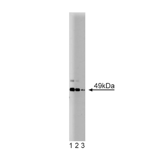
Western blot analysis for JNK/SAPK (pT183/pY185). HeLa cells (Human cervical epitheloid carcinoma; ATCC CCL-2) were either left untreated (lane 1) or treated with 25 µg/mL anisomycin, an antibiotic and protein synthesis inhibitor, for 15 min at 37°C (lane 2). The top panel was probed with a mouse anti-JNK/SAPK1 antibody (Cat. No. 610627) and the bottom panel was probed with the mouse anti-JNK/SAPK1 (pT183/pY185) antibody at a 1:250 dilution with bands observable at ~ 43 kDa & ~ 56 kDa.

Western blot analysis for JNK/SAPK (pT183/pY185). HeLa cells (Human cervical epitheloid carcinoma; ATCC CCL-2) were either left untreated (lane 1) or treated with 25 µg/mL anisomycin, an antibiotic and protein synthesis inhibitor, for 15 min at 37°C (lane 2). The top panel was probed with a mouse anti-JNK/SAPK1 antibody (Cat. No. 610627) and the bottom panel was probed with the mouse anti-JNK/SAPK1 (pT183/pY185) antibody at a 1:250 dilution with bands observable at ~ 43 kDa & ~ 56 kDa.
Preparation And Storage
Recommended Assay Procedures
Western blot: Please refer to http://www.bdbiosciences.com/pharmingen/protocols/Western_Blotting.shtml
Product Notices
- Since applications vary, each investigator should titrate the reagent to obtain optimal results.
- Please refer to www.bdbiosciences.com/us/s/resources for technical protocols.
- Caution: Sodium azide yields highly toxic hydrazoic acid under acidic conditions. Dilute azide compounds in running water before discarding to avoid accumulation of potentially explosive deposits in plumbing.
- Source of all serum proteins is from USDA inspected abattoirs located in the United States.
Companion Products

The Ras signaling pathway links the signals from growth factor receptors with the activation of the MAPK kinase cascade of phosphorylation leading to cell growth and differentiation. External stimuli, like endotoxins, UV irradiation, heat, and hyperosmolarity, induce an array of cellular responses that culminate with gene expression, ultimately dictating an adaptation to the new environment. Small GTPases of the Rho family, including cdc42, Rac1, and Rho, transmit the stress signals that initiate the signal cascade. JNK is a c-Jun kinase that was also identified as SAPK1 and MAPKp49. JNK/SAPK, along with p38 and RK5/BMK1, comprise three classes of stress-activated MAPK groups. Complete activation of JNK/SAPK requires the phosphorylation of both Thr183 and Tyr185, which are located in a Thr-X-Tyr motif. The activation of these residues is believed to be carried out by MKK4 and MKK7. Active JNK/SAPK phosphorylates other kinases and multiple transcription factors that induce expression of genes, such as proinflammatory cytokines.
Development References (4)
-
Fleming Y, Armstrong CG, Morrice N, Paterson A, Goedert M, Cohen P. Synergistic activation of stress-activated protein kinase 1/c-Jun N-terminal kinase (SAPK1/JNK) isoforms by mitogen-activated protein kinase kinase 4 (MKK4) and MKK7. Biochem J. 2000; 352:145-154. (Biology). View Reference
-
Hinton DR, Henderson VW, Blanks JC, Rudnicka M, Miller CA. Monoclonal antibodies react with neuronal subpopulations in the human nervous system. J Comp Neurol. 1988; 267(3):398-408. (Biology). View Reference
-
Kyriakis JM, Avruch J. Mammalian mitogen-activated protein kinase signal transduction pathways activated by stress and inflammation. Physiol Rev. 2001; 81(2):807-869. (Biology). View Reference
-
Mohit AA, Martin JH, Miller CA. p493F12 kinase: a novel MAP kinase expressed in a subset of neurons in the human nervous system. Neuron. 2001; 14(1):76-78. (Biology). View Reference
Please refer to Support Documents for Quality Certificates
Global - Refer to manufacturer's instructions for use and related User Manuals and Technical data sheets before using this products as described
Comparisons, where applicable, are made against older BD Technology, manual methods or are general performance claims. Comparisons are not made against non-BD technologies, unless otherwise noted.
For Research Use Only. Not for use in diagnostic or therapeutic procedures.