
Overview
The capabilities of flow cytometry instruments in terms of available lasers, detectors and number of measurable parameters have expanded tremendously over the years. Concomitant with that, there is an exponential increase in the number of fluorochromes available for flow cytometry, extending the limits of possibilities for new discoveries. Conventional flow cytometry collects only a discrete portion of the emission spectrum as defined by a single filter per fluorochrome, whereas spectral flow cytometry collects the full spectrum. This increases the flexibility and the capability of spectral flow by enabling autofluorescence profiling and extraction, detection of more fluorochromes per laser line and increased panel size. Find out how spectral flow cytometry differs from conventional flow cytometry and how to design your panels for spectral flow cytometry.
Spectral vs conventional flow cytometry comparison
| Feature | Spectral | Conventional |
| Detector and fluorochrome ratio | More detectors than fluorochromes | 1:1 |
| Individual fluorochrome contribution determination | Unmixing | Compensation |
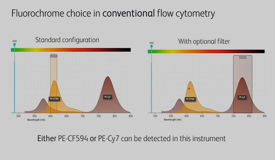
| Fluorochrome choice | Dependent on laser configuration | Dependent on laser and filter configuration |
| Autofluorescence extraction | Yes | No |
| Resolution of highly similar fluorochromes | Yes | No |
| Multi-color panel capability | 40+ colors | ~28 colors |
What remains the same?
- Workflow (sample prep, staining, acquisition)
- Need for accurate single-stain controls for unmixing
- Panel design principles
Introduction to spectral flow cytometry
Watch the spectral flow cytometry videos below to explore various topics, such as the fundamentals of spectral and conventional flow cytometry, how you can use more fluorochromes per laser line using spectral flow cytometry, how to use spectral flow cytometry for increased panel size, and more.
Fundamentals of spectral versus conventional flow cytometry
The video addresses the following questions:
- What is spectral flow cytometry?
- What are the similarities and differences between conventional and spectral flow cytometry?
- How do you read spectral signatures?
- What is unique about spectral flow cytometry?
Autofluorescence unmixing
The video addresses the following questions:
- What is autofluorescence unmixing?
- How can autofluorescence impact biological resolution?
- What are cell-specific autofluorescence signatures?
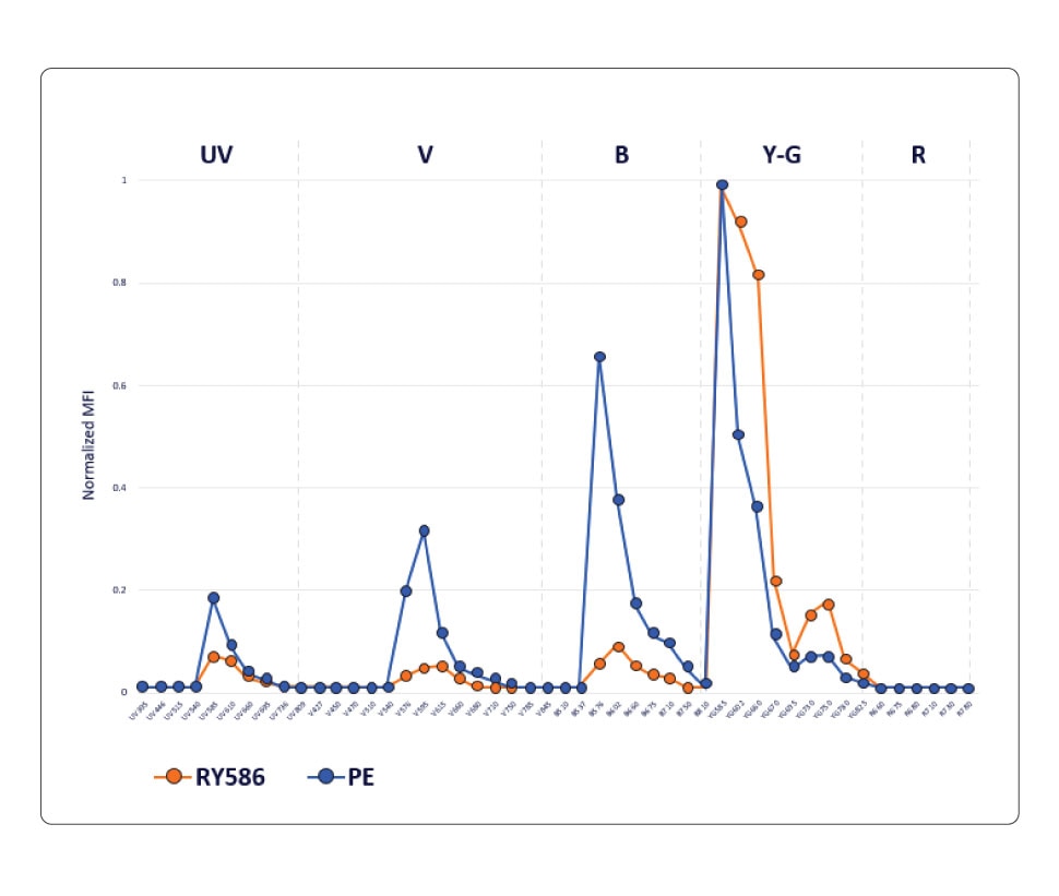
Fluorochrome choice flexibility
The video addresses the following questions:
- How to select fluorochromes in either conventional or spectral flow cytometry?
- How does spectral flow cytometry support the use of more fluorochromes per laser line?
- What are the advantages of using spectral flow cytometry for fluorescent protein detection?
Increased panel size
The video addresses the following questions:
- How does spectral flow cytometry enable resolution of highly similar fluorochromes?
- What are the similarity index and guidelines for the use of highly similar fluorochromes?
- What are the specifics behind a 40-color multicolor spectral flow cytometry panel?
Tools and considerations for the design of multicolor spectral flow cytometry
Spectral flow cytometry is an established technology that has increased the power, flexibility and complexity of flow cytometry. This webinar addresses the fundamental similarities and differences between conventional and spectral flow cytometry and dives deeper into facts and misconceptions around the principles and tools for multicolor spectral flow cytometry panel design.
Watch the webinar
- What is spectral flow cytometry and what makes it unique
- Fundamental rules and tools for panel design common to conventional and spectral flow cytometry
- The do’s and don’ts of panel design specific to spectral flow cytometry
BD Horizon RealYellow™ and BD Horizon RealBlue™ Reagents
Knowing which fluorochrome to use for each marker in your panel and assigning them in a way that can lead to deeper biological insights can sometimes be difficult. BD Biosciences offers new panel design resources to help with these choices as well as a wide array of antibody specificities and fluorochromes for flexible and easy panel design. We’re also developing new reagents such as our family of BD Horizon RealYellow™ and BD Horizon RealBlue™ Reagents with optimized emission spectra and reduced cross-laser excitation. Our goal is to help build your confidence to attain dependable data and streamline the path to accurate results.

BD Biosciences offers cell analyzers and sorters that support both conventional and spectral flow cytometry
Spectral flow cytometry represents an alternative data acquisition and analysis strategy to conventional flow cytometry providing advantages in flexibility of fluorescent label inputs without having to change filters, as well as the ability to multiplex more fluorescent labels in one multicolor sample.
Increase your experimental capabilities
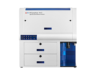
This 40-color panel, created on the BD FACSymphony™ A5 SE Cell Analyzer, uses carefully selected fluorescence reagents to characterize a variety of immune cell populations in human peripheral blood.
Get more information from the BD FACSymphony™ A5 SE Cell Analyzer brochure.
The BD FACSymphony™ A5 SE Cell Analyzer features five fixed-alignment lasers and 48 detectors for maximum coverage of the fluorochrome emission spectrum. By enabling both spectral unmixing and compensation-based workflows, the BD FACSymphony™ A5 SE Cell Analyzer increases flexibility in fluorochrome choices and permits simultaneous analysis of fluorochromes with similar spectral signatures. Spectral unmixing can also be used to resolve critical cell populations using autofluorescence unmixing.
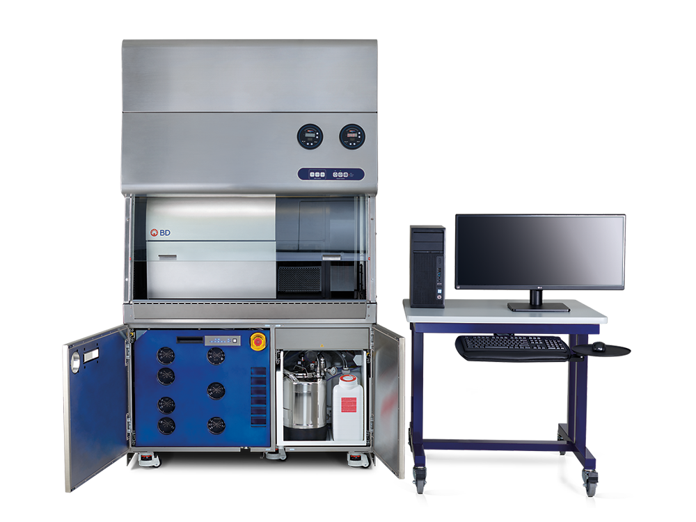
The BD FACSymphony™ S6 Cell Sorter builds on the foundations of the BD FACSymphony™ A5 Cell Analyzer with easy migration of optimized panels from analyzer to sorter. The BD FACSymphony™ S6 Cell Sorter supports both conventional compensation and spectral unmixing methods, with flexibility for up to nine spatially separated lasers and 50 parameters. Together with bioinformatics analysis tools and the broad portfolio of BD high-parameter reagents, the BD FACSymphony™ S6 Cell Sorter provides an end-to-end solution for high-parameter experimental success.
For Research Use Only. Not for use in diagnostic or therapeutic procedures.
Alexa Fluor (AF) is a trademark of Thermo Fisher Scientific. Cy is a trademark of Global Life Sciences Germany GmbH or an affiliate doing business as Cytiva.