





Waters Biosciences (formerly BD Biosciences) has led advancements in flow cytometry for more than 50 years, driving scientific discoveries and improving lives. Our high-performance flow cytometers, cell analyzers, and sorters, paired with intuitive software and world-class support deliver precise, reproducible results.
Our clinical cell analyzers deliver workflow automation, standardization across instruments and sites, enabling precise, reproducible results for clinical applications, while offering LIS connectivity and remote access.
Our latest research cell analyzers and sorters build on innovations in spectral flow cytometry and real-time, image-enabled spectral analysis and sorting. These technologies provide unparalleled experimental power and streamlined workflows, enabling consistent results while revealing single-cell insights that were previously undetectable.
BD FACSDiscover™ Flow Cytometers
Advanced High-Parameter Spectral Cell Analyzers and Sorters (For research use only)
Enhance your experimental power with advanced spectral cell analysis and integrated real-time imaging, leveraging breakthrough technologies to enable deeper characterization of cellular heterogeneity and functional biology.
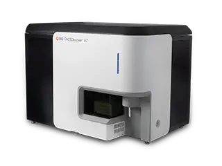
BD FACSDiscover™ A7 Cell Analyzer
50+ Color Spectral Cell Analyzer (RUO)
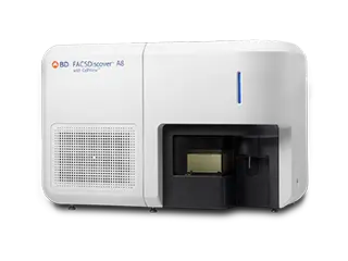
BD FACSDiscover™ A8 Cell Analyzer
50+ Color Spectral Cell Analyzer with Real-Time Imaging (RUO)
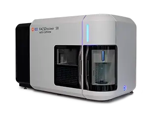
BD FACSDiscover™ S8 Cell Sorter
50+ Color Spectral Cell Sorter with Real-Time Imaging (RUO)
BD FACSLyric™ and BD FACSCanto™ Clinical Flow Cytometers
(For In Vitro Diagnostic Use)
With the BD FACSLyric™ Clinical Flow Cytometry System, discover a flow cytometer engineered to advance your laboratory workflow through high‑performance detection, robust automation, and rigorous standardization—delivering superior sensitivity and enhanced resolution.
With the BD FACSCanto™ Clinical Flow Cytometry System, experience the reliability of performance, accuracy and ease of use necessary for your clinical laboratory.

12-Color Conventional Cell Analyzer (IVD)
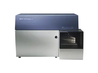
8-Color Conventional Cell Analyzer (IVD)
BD FACSymphony™ Flow Cytometers
Customizable Flow Cytometers for high-parameter analysis (For Research Use Only)
Explore the BD FACSymphony™ Cell Analyzers, which deliver flexible, high parameter flow cytometry with advanced sensitivity and a familiar BD FACSDiva™ Software workflow.
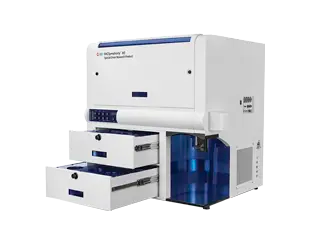
30-Color Conventional Cell Analyzer (RUO)
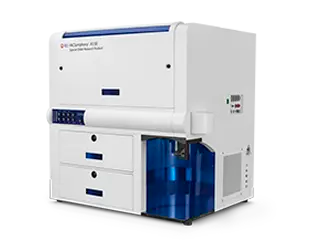
34+ Color Spectral Cell Analyzer (RUO)
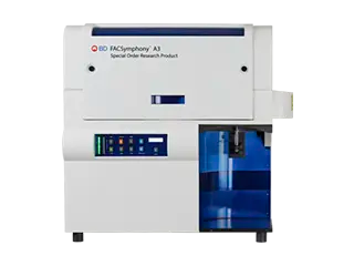
28-Color Conventional Cell Analyzer (RUO)
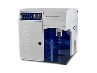
16-Color Conventional Cell Analyzer (RUO)
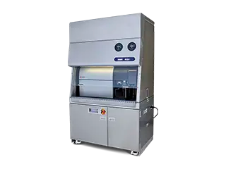
28-Color Conventional Cell Sorter (RUO)
BD LSR Fortessa™Flow Cytometers
Special order cell analyzers for core labs (For Research Use Only)
Leverage this core lab workhorse that provides power, performance, and consistency to core labs.
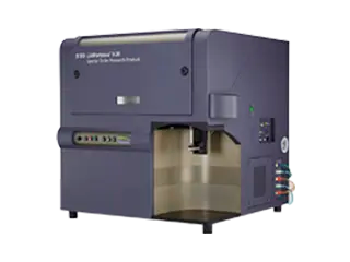
5-Laser, 18-Color Conventional Cell Analyzer (RUO)
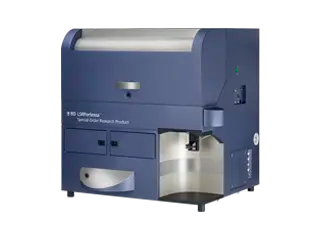
4-Laser, 18-Color Conventional Cell Analyzer (RUO)
BD FACSAria™ Flow Cytometers
Dependable cell sorters with multicolor performance (For Research Use Only)
Leverage the high sensitivity, multicolor performance, ease of use, and biosafety of these legendary cell sorters.
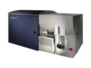
18-Color Conventional Cell Sorter (RUO)
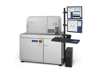
BD FACSAria™ Fusion Cell Sorter
18-Color Conventional Cell Sorter with Integrated Biosafety Cabinet (RUO)
Benchtop, Compact Cell Analyzers and Sorters
(For Research Use Only)
With the BD FACSCelesta™ Cell Analyzer, make multicolor flow cytometry accessible with this benchtop cell analyzer with the ability to detect and analyze up to 14 parameters in a single sample.
Utilize the compact, lightweight BD® Accuri C6 Plus Personal Flow Cytometer with the operational simplicity and analytical power of advanced flow cytometers.
With the BD FACSMelody™ Cell Sorter, discover a cost-effective and easy-to-use cell sorter with advanced automation technology.

12-Color Conventional Cell Analyzer (RUO)

BD® Accuri C6 Plus Personal Flow Cytometer
4-Color Conventional Cell Analyzer (RUO)

9-Color Conventional Cell Sorter (RUO)
Clinical Cell Analyzers
Select Your Clinical Cell Analyzer
Learn the specifications of each of our clinical cell analyzer and choose the best that suits your needs.
| Instrument | Parameters (up to) | Colors (up to) | Lasers (up to) | Software | Regulatory Status | Recommended Sample Prep System | LIS Integration Capability | Sample Loader | Loader Options |
| BD FACSLyric™ | 14 | 12 | 3 | FACSuite™ Clinical Software | US-IVD Up to 6 colors | FACSDuet | BD FACS™ Workflow Manager | Plates & Tubes | |
| BD FACSCanto™ | 10 | 8 | 3 | FACSCanto Clinical and FACSDiva Software | US-IVD Up to 6 colors | BD FACS™ SPA III/LWA | BD FACS™ Workflow Manager | Tubes |
Learn more on how we support our discontinued FACSCalibur™ and FACSCount™ instruments. Find out more information on ourFACSVia™ System.
Research Cell Analyzers
Select Your Research Cell Analyzer
Explore our expansive suite of research cell analyzers and select the best one that works for you.
| Instruments | Detectors (up to) | Colors (up to) | Lasers (up to) | Customizable | Benchtop | Software | Loader Options |
| 81 | 50+ | 5 | N | Y | BD FACSChorus™ | Plates & Custom Tube Racks | |
| BD FACSDiscover™ A8 | 86 | 50+ | 5 | BD FACSChorus™ | Plates & Custom Tube Racks | ||
| BD FACSymphony™ A1 | 19 | 16 | 4 | Inquire | BD FACSDiva™ | Plates | |
| FACSymphony™ A3 | 30 | 28 | 5 | BD FACSDiva™ | Plates | ||
| FACSymphony™ A5 | 50 | 30 | 9 | BD FACSDiva™ | Plates | ||
| FACSymphony™ A5 SE | 50 | 34+ | 5 | BD FACSDiva™ | Plates | ||
| BD LSRFortessa™ X-20 | 20 | 18 | 5 | BD FACSDiva™ | Plates | ||
| BD LSRFortessa™ | 20 | 18 | 5 | BD FACSDiva™ | Plates | ||
| BD Accuri™ C6 Plus | 6 | 4 | 2 | BD Accuri™ C6 Plus | Plates & Tubes | ||
| BD FACSCelesta™ | 14 | 12 | 3 | BD FACSDiva™ | Plates |
Find more information on the discontinued FACSVerse™ Cell Analyzer and explore how to trade in your BD™ LSRII Flow Cytometer
Research Cell Sorters
Select Your Research Cell Sorter
| Instruments | Detectors (up to) | Imaging Detectors | Colors (up to) | Lasers (up to) | Laser Options | Multiway sorting | Software | Customizable | Nozzle Size (µm) | Automated Sort Setup & Guided Workflow |
| BD FACSDiscover™ S8 | 86 | 6 | N/A | 5 | 5 | 6-way | BD FACSChorus™ | 85, 100, 130 | ||
| BD FACSymphony™ S6 | 50 | 0 | 28 | 9 | >25* | 6-way | BD FACSDiva™ | 70, 85, 100, 130 | ||
| BD FACSMelody™ | 11 | 0 | 9 | 3 | 4 | 4-way | BD FACSChorus™ | 100 | ||
| BD FACSAria™ III | 20 | 0 | 18 | 4 | 5 | 4-way | BD FACSDiva™ | 70, 85, 100, 130 | ||
| BD FACSAria™ Fusion | 20 | 0 | 18 | 5 | 5* | 4-way | BD FACSDiva™ | 70, 85, 100, 130 |
*More laser choices and configurations are available through Special Order Research (SORP).
Find the consumables and resources for the BD Influx™ and BD FACSJazz™ Cell Sorters
Flow Cytometry Training Materials
Explore our comprehensive training resources, including introductory materials on the fundamentals of flow cytometry and advanced guides with detailed instructions and videos for specific flow cytometer models. Access both basic and product-specific training:

FAQs
- What is a flow cytometer and what are the differences between a cell analyzer and a cell sorter?
Flow cytometers are specialized instruments that measure distinct characteristics of cells as they pass through a laser beam. By recording both light scatter and fluorescent signals emitted by cell-bound fluorochromes, flow cytometers support applications spanning basic cell analysis to immunophenotyping of complex cell populations. Flow cytometers can be divided between cell analyzers and cell sorters. While analyzers are built to support faster workflows and higher throughput between samples, cell sorters take flow cytometry one step further by isolating specific cell populations and dispensing them into tubes or microplates for downstream use, rather than simply discarding the sample. The sorted cells can then be used for applications including functional assays, 3D cell culture, and antibody generation, as well as genomics, sequencing, and imaging studies.
- How is traditional flow cytometry distinct from spectral flow cytometry?
Traditional (polychromatic) flow cytometers measure a discrete portion of each fluorochrome's emission spectrum, determined by optical filters. Spectral flow cytometers instead measure the full emission spectrum of each fluorochrome, often referred to as a spectral signature. An important advantage of spectral flow cytometry is that it allows for using fluorochromes with similar emission profiles in the same experiment, increasing the number of parameters that can be simultaneously detected. Additionally, with spectral flow cytometry, autofluorescence can be treated as separate measurements and accounted for as controls to be used for unmixing. This process, commonly referred to as “autofluorescence extraction” may support reducing background signals and thus improving the resolution. Discover the spectrally-enabled BD FACSDiscover™ S8 Cell Sorter
- How does adding imaging to flow cytometry help to advance scientific research?
The introduction of real-time imaging capabilities to spectral flow cytometers has further expanded their utility, enabling higher parameter analysis that can unlock deeper biological insights. Specifically, by combining common flow cytometry parameters with morphological and spatial information not captured by traditional instruments, imaging flow cytometry can help to resolve previously unanswerable questions. Additionally, imaging has the potential to improve sort performance, including through better discrimination of large or irregularly shaped cells (e.g., dendritic cells) from doublets or aggregates. Experience real-time imaging with the BD FACSDiscover™ A8 Cell Analyzer and the BD FACSDiscover™ S8 Cell Sorter
- How many parameters can be simultaneously measured on a flow cytometer?
With traditional flow cytometry, it is common for researchers to simultaneously detect 8-18 different parameters. However, with careful panel design, it is now possible to measure up to 30 markers with instruments such as the BD FACSymphony™ A5 Cell Analyzer. Spectral flow cytometry enables more markers to be detected in the same experiment, with instruments such as the BD FACSymphony™ S6 Cell Sorter and BD FACSDiscover™ S8 Cell Sorter allowing as many as 50 markers to be measured.
- What is the maximum event rate for flow cytometry?
The event rate, defined as the number of single particles detected per second, is dependent on the flow cytometer’s fluidics and electronics systems, which differ between technologies. The fluidics system, including the sample injection method used (pressurized, syringe-pumped or vacuum based), determines the flow rate, measured as mL/min. The electronics system is responsible for the flow cytometer’s processing capacity and depends on the number of detectable parameters and calculations it is capable of handling during data acquisition (acquisition rate, measured as events/sec). Within Waters Biosciences’ (formerly BD Biosciences) portfolio, instruments are capable of handling up to 70,000 events per second and sorting up to 25,000 events per second. However, while these high event rates are achievable, it is generally recommended to run samples at lower rates for greater consistency and precision across acquired events (lower variation). For instance, a sample prepared at 1 x 10⁶ cells/mL and suspended on 0.3 mL can be acquired at a 5,000 events/second rate using average flow rates (~ 60 mL/min). In this example, by controlling the flow rate during acquisition (lowering or increasing sample intake), the event rate can be adjusted to a range between 2,500 and 10,000 events/second so that it suits user preferences or specific application needs.
- What types of samples can be analyzed using a flow cytometer?
Flow cytometry has broad utility for evaluating particles in liquid suspensions, provided the particles fall within the specific size limits of the instrument being used. The main applications of flow cytometry are related to analyzing specialized cells from multicellular organisms, or unicellular organisms such as bacteria and algae. However, flow cytometry can also be used to detect and quantify soluble analytes secreted by cells, such as cytokines and chemokines, when captured by synthetic beads; to evaluate and sort pollen grains; and to study extracellular vesicles (EVs) including exosomes, microvesicles, and apoptotic bodies. Despite this diversity, the most common sample types for flow cytometry are cells obtained from biological material including whole blood, bone marrow, and solid tissues.
- What sample volume and cell concentration is required for flow cytometry?
The sample volume and cell concentration required for flow cytometry depends on the type of sample being used and the experimental objectives. As a general rule, samples are traditionally prepared as suspensions between 0.2 and 0.5 mL at a concentration of 0.5 – 1 x 10⁶ cells/mL. but these values should always be optimized for your flow cytometer and experimental model. For limited samples, instruments such as the BD FACSDiscover A8 Cell Analyzer have a minimum sample requirement of just 3 mL for samples processed on 96-well plates and 12x75mm Falcon® tubes (minimum acquisition volume of 2uL plus a dead volume of 1uL).
- What are spillover, compensation, and spectral unmixing?
Spillover occurs whenever the fluorescence emission of one fluorochrome is captured by a detector designed to measure signal from another fluorochrome. To correct for spillover, single-color controls must be acquired to determine a reference for positive signals from every fluorochrome used in a given experiment. Similarly, negative controls that lack any of the fluorochromes being used should be acquired from an unstained sample. Conventional flow cytometers, which use one detector per fluorochrome, apply the information collected from the controls to a mathematical process called ‘compensation’, such that each detector only reports the signal coming from its intended fluorochrome. Spectral flow cytometers instead measure the full emission spectrum across many detectors and use a different type of math called 'spectral unmixing' to determine how much signal came from each fluorochrome in the experiment, based on spectral signatures.
BD Flow Cytometers are Class 1Laser Products. The BD FACSCanto™ Flow Cytometer is for In Vitro Diagnostic Use for up to six colors. Seven to ten colors are for Research Use Only.
The BD FACSLyric™ Flow Cytometer is for In Vitro Diagnostic Use with BD FACSuite™ Clinical Application for up to six colors. The BD FACSLyric™ Flow Cytometer is for Research Use Only with BD FACSuite™ Application for up to 12 colors. Not for use in diagnostic or therapeutic procedures.
BD FACSDiscover™ A8 Cell Analyzer, BD FACSDiscover™ S8 Cell Sorter, BDFACSymphony™ A5 Cell Analyzer, BDFACSymphony™ A5 SE Cell Analyzer, BDFACSymphony™ A3 Cell Analyzer, BDFACSymphony™ A1 Cell Analyzer, BDFACSymphony™ S6 Cell Sorter, BD LSRFortessa™ Cell Analyzer, BD LSRFortessa™ X-20 Cell Analyzer, BD FACSAria™ III Cell Sorter, BD FACSAria™ Fusion Cell Sorter, BD FACSMelody™ Flow Cytometer, BD® Accuri C6 Plus Personal Flow Cytometer and BD FACSCelesta™ Flow Cytometer are for Research Use Only. Not for use in diagnostic or therapeutic procedures.
In the EU, the BD FACSLyric™ Flow Cytometer with the BD FACSuite™ Clinical and BD FACSuite™ Applications are an in vitro diagnostic medical device bearing a CE mark.