-
Reagents
- Flow Cytometry Reagents
-
Western Blotting and Molecular Reagents
- Immunoassay Reagents
-
Single-Cell Multiomics Reagents
- BD® OMICS-Guard Sample Preservation Buffer
- BD® AbSeq Assay
- BD® Single-Cell Multiplexing Kit
- BD Rhapsody™ ATAC-Seq Assays
- BD Rhapsody™ Whole Transcriptome Analysis (WTA) Amplification Kit
- BD Rhapsody™ TCR/BCR Next Multiomic Assays
- BD Rhapsody™ Targeted mRNA Kits
- BD Rhapsody™ Accessory Kits
- BD® OMICS-One Protein Panels
-
Functional Assays
-
Microscopy and Imaging Reagents
-
Cell Preparation and Separation Reagents
Old Browser
Looks like you're visiting us from {countryName}.
Would you like to stay on the current location site or be switched to your location?
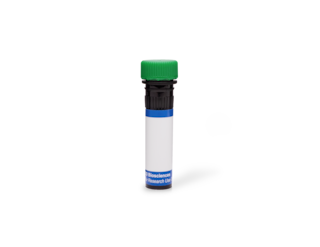
BD Transduction Laboratories™ Purified Mouse Anti-Paxillin
Clone 349/Paxillin (RUO)


Western blot analysis of Paxillin on a A431 lysate. Lane 1: 1:10,000; lane 2: 1:20,000; lane 3: 1:40,000 dilution of the anti- Paxillin antibody.


Immunofluoresence staining of human fibroblasts.


Regulatory Status Legend
Any use of products other than the permitted use without the express written authorization of Becton, Dickinson and Company is strictly prohibited.
Preparation And Storage
Product Notices
- Since applications vary, each investigator should titrate the reagent to obtain optimal results.
- Please refer to www.bdbiosciences.com/us/s/resources for technical protocols.
- Caution: Sodium azide yields highly toxic hydrazoic acid under acidic conditions. Dilute azide compounds in running water before discarding to avoid accumulation of potentially explosive deposits in plumbing.
- Source of all serum proteins is from USDA inspected abattoirs located in the United States.
Companion Products


A number of cytoskeletal proteins are tyrosine phosphorylated in Rous sarcoma virus-transformed chick embryo fibroblasts. One of these is the 68 kDa paxillin protein. Paxillin is a cytoskeletal component that localizes to the focal adhesions at the ends of actin stress fibers. It is also present in the focal adhesions of Madin-Darby canine kidney epithelial cells, but is absent from the cell adherens junctions of these cells. Paxillin purified from chicken gizzard migrates as a diffuse band on SDS-PAGE with molecular weight of 65-70 kDa. It binds to the rod domain of vinculin, another focal adhesion protein. It is thought that phosphorylation of paxillin may have a role in that disassembly of focal adhesions and stress fibers during transformation.
This antibody is routinely tested by Western blot analysis. Other applications were tested at BD Biosciences Pharmingen during antibody development only or reported in the literature.
Development References (5)
-
Even-Ram SC, Maoz M, Pokroy E, et al. Tumor cell invasion is promoted by activation of protease activated receptor-1 in cooperation with the alpha vbeta 5 integrin. J Biol Chem. 2001; 276(14):10952-10962. (Biology: Immunofluorescence, Immunoprecipitation, Western blot). View Reference
-
Glenney JR Jr, Zokas L. Novel tyrosine kinase substrates from Rous sarcoma virus-transformed cells are present in the membrane skeleton. J Cell Biol. 1989; 108(6):2401-2408. (Biology). View Reference
-
Nikolopoulos SN, Turner CE. Actopaxin, a new focal adhesion protein that binds paxillin LD motifs and actin and regulates cell adhesion. J Cell Biol. 2000; 151(7):1435-1447. (Biology: Immunofluorescence, Western blot). View Reference
-
Wine RN, Chapin RE. Adhesion and signaling proteins spatiotemporally associated with spermiation in the rat. J Androl. 1999; 20(2):198-213. (Biology: Immunohistochemistry, Western blot). View Reference
-
van de Water B, Houtepen F, Huigsloot M, Tijdens IB. Suppression of chemically induced apoptosis but not necrosis of renal proximal tubular epithelial (LLC-PK1) cells by focal adhesion kinase (FAK). Role of FAK in maintaining focal adhesion organization after acute renal cell injury. J Biol Chem. 2001; 276(29):36183-36193. (Biology: Immunofluorescence, Western blot). View Reference
Please refer to Support Documents for Quality Certificates
Global - Refer to manufacturer's instructions for use and related User Manuals and Technical data sheets before using this products as described
Comparisons, where applicable, are made against older BD Technology, manual methods or are general performance claims. Comparisons are not made against non-BD technologies, unless otherwise noted.
For Research Use Only. Not for use in diagnostic or therapeutic procedures.
Refer to manufacturer's instructions for use and related User Manuals and Technical Data Sheets before using this product as described.
Comparisons, where applicable, are made against older BD technology, manual methods or are general performance claims. Comparisons are not made against non-BD technologies, unless otherwise noted.