-
Reagents
- Flow Cytometry Reagents
-
Western Blotting and Molecular Reagents
- Immunoassay Reagents
-
Single-Cell Multiomics Reagents
- BD® OMICS-Guard Sample Preservation Buffer
- BD® AbSeq Assay
- BD® Single-Cell Multiplexing Kit
- BD Rhapsody™ ATAC-Seq Assays
- BD Rhapsody™ Whole Transcriptome Analysis (WTA) Amplification Kit
- BD Rhapsody™ TCR/BCR Next Multiomic Assays
- BD Rhapsody™ Targeted mRNA Kits
- BD Rhapsody™ Accessory Kits
- BD® OMICS-One Protein Panels
-
Functional Assays
-
Microscopy and Imaging Reagents
-
Cell Preparation and Separation Reagents
-
- BD® OMICS-Guard Sample Preservation Buffer
- BD® AbSeq Assay
- BD® Single-Cell Multiplexing Kit
- BD Rhapsody™ ATAC-Seq Assays
- BD Rhapsody™ Whole Transcriptome Analysis (WTA) Amplification Kit
- BD Rhapsody™ TCR/BCR Next Multiomic Assays
- BD Rhapsody™ Targeted mRNA Kits
- BD Rhapsody™ Accessory Kits
- BD® OMICS-One Protein Panels
- New Zealand (English)
-
Change country/language
Old Browser
Looks like you're visiting us from United States.
Would you like to stay on the current country site or be switched to your country?
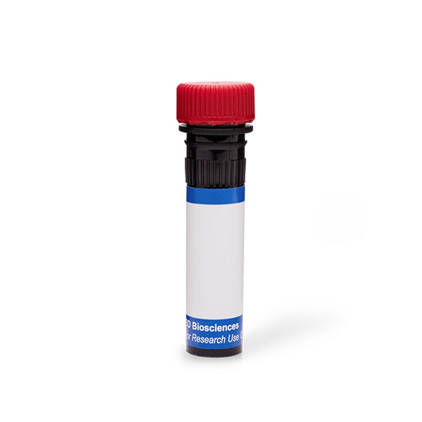
BD Pharmingen™ PE Mouse Anti- IRF4
Clone Q9-343 (RUO)

Flow cytometric analysis of IRF4 expression in human and mouse leucocytes. Human peripheral blood mononuclear cells (PBMC) were not stimulated (Top Left Plot) or stimulated (Top Right Plot) with phytohemagglutinin (PHA; 3 days). C57BL/6 mouse splenic B cells were not stimulated (Bottom Left Plot) or stimulated (Bottom Right Plot) with lipopolysaccharide (LPS; 2 days). PBMC were then fixed with BD Cytofix™ Fixation Buffer (Cat. No. 554655) and permeabilized and stained in BD Perm/Wash™ Buffer (Cat. No. 554723) with either PE Mouse IgG1, κ Isotype Control (Cat. No. 554680; dashed line histograms) or PE Mouse Anti-IRF4 antibody (Cat. No. Cat. No. 566646/566649; solid line histograms) at 0.25 µg/test. Mouse cells were similarly fixed, permeabilized, and stained using the BD Pharmingen™ Transcription Factor Buffer Set (Cat. No. 562574/562725). Histograms showing IRF4 expression (or Ig Isotype control staining) were derived from gated events with the forward and side light-scatter characteristics of intact leucocyte populations. Flow cytometric analysis was performed using a BD FACSCanto™ II System. Data shown on this Technical Data Sheet are not lot specific.

Flow cytometric analysis of IRF4 expression in human and mouse leucocytes. Human peripheral blood mononuclear cells (PBMC) were not stimulated (Top Left Plot) or stimulated (Top Right Plot) with phytohemagglutinin (PHA; 3 days). C57BL/6 mouse splenic B cells were not stimulated (Bottom Left Plot) or stimulated (Bottom Right Plot) with lipopolysaccharide (LPS; 2 days). PBMC were then fixed with BD Cytofix™ Fixation Buffer (Cat. No. 554655) and permeabilized and stained in BD Perm/Wash™ Buffer (Cat. No. 554723) with either PE Mouse IgG1, κ Isotype Control (Cat. No. 554680; dashed line histograms) or PE Mouse Anti-IRF4 antibody (Cat. No. Cat. No. 566646/566649; solid line histograms) at 0.25 µg/test. Mouse cells were similarly fixed, permeabilized, and stained using the BD Pharmingen™ Transcription Factor Buffer Set (Cat. No. 562574/562725). Histograms showing IRF4 expression (or Ig Isotype control staining) were derived from gated events with the forward and side light-scatter characteristics of intact leucocyte populations. Flow cytometric analysis was performed using a BD FACSCanto™ II System. Data shown on this Technical Data Sheet are not lot specific.


Flow cytometric analysis of IRF4 expression in human and mouse leucocytes. Human peripheral blood mononuclear cells (PBMC) were not stimulated (Top Left Plot) or stimulated (Top Right Plot) with phytohemagglutinin (PHA; 3 days). C57BL/6 mouse splenic B cells were not stimulated (Bottom Left Plot) or stimulated (Bottom Right Plot) with lipopolysaccharide (LPS; 2 days). PBMC were then fixed with BD Cytofix™ Fixation Buffer (Cat. No. 554655) and permeabilized and stained in BD Perm/Wash™ Buffer (Cat. No. 554723) with either PE Mouse IgG1, κ Isotype Control (Cat. No. 554680; dashed line histograms) or PE Mouse Anti-IRF4 antibody (Cat. No. Cat. No. 566646/566649; solid line histograms) at 0.25 µg/test. Mouse cells were similarly fixed, permeabilized, and stained using the BD Pharmingen™ Transcription Factor Buffer Set (Cat. No. 562574/562725). Histograms showing IRF4 expression (or Ig Isotype control staining) were derived from gated events with the forward and side light-scatter characteristics of intact leucocyte populations. Flow cytometric analysis was performed using a BD FACSCanto™ II System. Data shown on this Technical Data Sheet are not lot specific.
Flow cytometric analysis of IRF4 expression in human and mouse leucocytes. Human peripheral blood mononuclear cells (PBMC) were not stimulated (Top Left Plot) or stimulated (Top Right Plot) with phytohemagglutinin (PHA; 3 days). C57BL/6 mouse splenic B cells were not stimulated (Bottom Left Plot) or stimulated (Bottom Right Plot) with lipopolysaccharide (LPS; 2 days). PBMC were then fixed with BD Cytofix™ Fixation Buffer (Cat. No. 554655) and permeabilized and stained in BD Perm/Wash™ Buffer (Cat. No. 554723) with either PE Mouse IgG1, κ Isotype Control (Cat. No. 554680; dashed line histograms) or PE Mouse Anti-IRF4 antibody (Cat. No. Cat. No. 566646/566649; solid line histograms) at 0.25 µg/test. Mouse cells were similarly fixed, permeabilized, and stained using the BD Pharmingen™ Transcription Factor Buffer Set (Cat. No. 562574/562725). Histograms showing IRF4 expression (or Ig Isotype control staining) were derived from gated events with the forward and side light-scatter characteristics of intact leucocyte populations. Flow cytometric analysis was performed using a BD FACSCanto™ II System. Data shown on this Technical Data Sheet are not lot specific.

Flow cytometric analysis of IRF4 expression in human and mouse leucocytes. Human peripheral blood mononuclear cells (PBMC) were not stimulated (Top Left Plot) or stimulated (Top Right Plot) with phytohemagglutinin (PHA; 3 days). C57BL/6 mouse splenic B cells were not stimulated (Bottom Left Plot) or stimulated (Bottom Right Plot) with lipopolysaccharide (LPS; 2 days). PBMC were then fixed with BD Cytofix™ Fixation Buffer (Cat. No. 554655) and permeabilized and stained in BD Perm/Wash™ Buffer (Cat. No. 554723) with either PE Mouse IgG1, κ Isotype Control (Cat. No. 554680; dashed line histograms) or PE Mouse Anti-IRF4 antibody (Cat. No. Cat. No. 566646/566649; solid line histograms) at 0.25 µg/test. Mouse cells were similarly fixed, permeabilized, and stained using the BD Pharmingen™ Transcription Factor Buffer Set (Cat. No. 562574/562725). Histograms showing IRF4 expression (or Ig Isotype control staining) were derived from gated events with the forward and side light-scatter characteristics of intact leucocyte populations. Flow cytometric analysis was performed using a BD FACSCanto™ II System. Data shown on this Technical Data Sheet are not lot specific.

Flow cytometric analysis of IRF4 expression in human and mouse leucocytes. Human peripheral blood mononuclear cells (PBMC) were not stimulated (Top Left Plot) or stimulated (Top Right Plot) with phytohemagglutinin (PHA; 3 days). C57BL/6 mouse splenic B cells were not stimulated (Bottom Left Plot) or stimulated (Bottom Right Plot) with lipopolysaccharide (LPS; 2 days). PBMC were then fixed with BD Cytofix™ Fixation Buffer (Cat. No. 554655) and permeabilized and stained in BD Perm/Wash™ Buffer (Cat. No. 554723) with either PE Mouse IgG1, κ Isotype Control (Cat. No. 554680; dashed line histograms) or PE Mouse Anti-IRF4 antibody (Cat. No. Cat. No. 566646/566649; solid line histograms) at 0.25 µg/test. Mouse cells were similarly fixed, permeabilized, and stained using the BD Pharmingen™ Transcription Factor Buffer Set (Cat. No. 562574/562725). Histograms showing IRF4 expression (or Ig Isotype control staining) were derived from gated events with the forward and side light-scatter characteristics of intact leucocyte populations. Flow cytometric analysis was performed using a BD FACSCanto™ II System. Data shown on this Technical Data Sheet are not lot specific.


ImageTitle~BD Pharmingen™ PE Mouse Anti- IRF4

ImageTitle~BD Pharmingen™ PE Mouse Anti- IRF4


ImageTitle~BD Pharmingen™ PE Mouse Anti- IRF4
Regulatory Status Legend
Any use of products other than the permitted use without the express written authorization of Becton, Dickinson and Company is strictly prohibited.
Preparation And Storage
Product Notices
- Since applications vary, each investigator should titrate the reagent to obtain optimal results.
- An isotype control should be used at the same concentration as the antibody of interest.
- Caution: Sodium azide yields highly toxic hydrazoic acid under acidic conditions. Dilute azide compounds in running water before discarding to avoid accumulation of potentially explosive deposits in plumbing.
- For fluorochrome spectra and suitable instrument settings, please refer to our Multicolor Flow Cytometry web page at www.bdbiosciences.com/colors.
- Species cross-reactivity detected in product development may not have been confirmed on every format and/or application.
- Please refer to www.bdbiosciences.com/us/s/resources for technical protocols.
Data Sheets
Companion Products


The Q9-343 monoclonal antibody specifically recognizes human and mouse Interferon Regulatory Factor 4 (IRF4 or IRF-4) which is also known as Lymphocyte specific interferon regulatory factor (LSIRF), Multiple myeloma oncogene 1 (MUM1), or PU.1 interaction partner (PIP). IRF4 belongs to the Interferon Regulatory Factor (IRF) family of transcription factors that includes nine members, IRF1-9. IRF4 has a conserved N-terminal DNA binding domain with a unique tryptophan pentad repeat. Its C-terminal regulatory domain regulates IRF4 activity and mediates interactions with other IRF proteins, transcription factors and co-factors. IRF4 plays essential roles in the regulation of innate and adaptive immune responses. IRF4 is widely expressed in leucocytes and is essential for the development, activation, differentiation, and/or apoptosis of T helper (Th) cell subsets including Th2, Th9, Th17, T follicular helper (Tfh) cells, or T (Treg) cells. IRF4 is involved in the development or differentiation of CD8+ effector and memory cells, B cells and plasma cells, as well as different dendritic cell (DC) subsets and M2 macrophages. IRF4 is also expressed by adipocytes and melanocytes. Cellular IRF4 expression is primarily upregulated by antigen-receptor engagement, or by stimulation with lipopolysaccharide (LPS), CD40, or IL-4, rather than by interferons. TLR4 has been implicated in suppressing or promoting oncogenisis and autoimmunity.

Development References (6)
-
Huber M, Lohoff M. IRF4 at the crossroads of effector T-cell fate decision.. Eur J Immunol. 2014; 44(7):1886-95. (Biology). View Reference
-
Nam S, Lim JS. Essential role of interferon regulatory factor 4 (IRF4) in immune cell development.. Arch Pharm Res. 2016; 39(11):1548-1555. (Biology). View Reference
-
Pernis AB. The role of IRF-4 in B and T cell activation and differentiation.. J Interferon Cytokine Res. 2002; 22(1):111-20. (Biology). View Reference
-
Shaffer AL, Emre NC, Romesser PB, Staudt LM. IRF4: Immunity. Malignancy! Therapy?. Clin Cancer Res. 2009; 15(9):2954-61. (Biology). View Reference
-
Yanai H, Negishi H, Taniguchi T. The IRF family of transcription factors: Inception, impact and implications in oncogenesis.. Oncoimmunology. 2012; 1(8):1376-1386. (Biology). View Reference
-
Zhao GN, Jiang DS, Li H. Interferon regulatory factors: at the crossroads of immunity, metabolism, and disease.. Biochim Biophys Acta. 2015; 1852(2):365-78. (Biology). View Reference
Please refer to Support Documents for Quality Certificates
Global - Refer to manufacturer's instructions for use and related User Manuals and Technical data sheets before using this products as described
Comparisons, where applicable, are made against older BD Technology, manual methods or are general performance claims. Comparisons are not made against non-BD technologies, unless otherwise noted.
For Research Use Only. Not for use in diagnostic or therapeutic procedures.
Refer to manufacturer's instructions for use and related User Manuals and Technical Data Sheets before using this product as described.
Comparisons, where applicable, are made against older BD technology, manual methods or are general performance claims. Comparisons are not made against non-BD technologies, unless otherwise noted.