Skip to main content Skip to navigation
PE Mouse Anti-Human IL-1α
Expression of IL-α by stimulated CD14+ human monocytes. Human PBMCs were stimulated for 6 hours with LPS (10 ng/ml final concentration) in the presence of GolgiStop™ (2 µM final concentration; Cat. No. 554724). The PBMC were stained with FITC-mouse anti-human CD14 antibody (Clone M5E2, Cat. No. 555397) fixed/permeabilized, and then stained with 0.25 µg of PE-mouse anti-human IL-1α antibody (PE-364-3B3-14, Cat. No. 554561), (left panel). The data reflects gating on monocytes, based on forward and side scatter. To demonstrate specificity of staining, the binding of PE-364-3B3-14 was blocked by preincubation of the PE-364-3B3-14 with a molar excess of recombinant human IL-1α (middle panel), and by the preincubation of the fixed/permeabilized cells with an excess of the unlabeled \"cold\" 364-3B3-14 mouse antibody (5 µg; Cat. No. 551223; right panel). The quadrant markers for the bivariate dot plots were set based on the fixed/permeabilized unstained cell controls, and verified with the recombinant cytokine and \"cold\" antibody specificity controls.
Product Details
Down Arrow Up Arrow


BD Pharmingen™
Human (QC Testing)
Mouse IgG1, κ
recombinant human IL-1α
Intracellular staining (flow cytometry) (Routinely Tested)
0.2 mg/ml
AB_395480
Aqueous buffered solution containing ≤0.09% sodium azide.
RUO


Preparation And Storage

Store undiluted at 4°C and protected from prolonged exposure to light. Do not freeze. The monoclonal antibody was purified from tissue culture supernatant or ascites by affinity chromatography. The antibody was conjugated with R-PE under optimum conditions, and unconjugated antibody and free PE were removed.

Recommended Assay Procedures

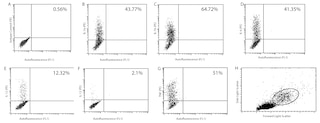
Immunofluorescent Staining and Flow Cytometric Analysis: The 364-3B3-14 antibody is useful for immunofluorescent staining and flow cytometric analysis to identify and enumerate IL-1α producing cells within mixed cell populations. The PE-conjugated 364-3B3-14 antibody is especially suitable for these studies (see figure). For optimal immunofluorescent staining and flow cytometric analysis, this anti-cytokine antibody should be titrated (≤ 0.5 µg mAb / 1X10^6 cells). For specific methodology, please refer to the resources/protocol section on our website: http://www.bdbiosciences.com/resources/index.jsp

Product Notices

  1. Since applications vary, each investigator should titrate the reagent to obtain optimal results.
  2. For fluorochrome spectra and suitable instrument settings, please refer to our Multicolor Flow Cytometry web page at www.bdbiosciences.com/colors.
  3. Caution: Sodium azide yields highly toxic hydrazoic acid under acidic conditions. Dilute azide compounds in running water before discarding to avoid accumulation of potentially explosive deposits in plumbing.
  4. This product is manufactured and sold under license from Pestka Biomedical Laboratories, Inc. (d/b/a PBL InterferonSource) and may be used solely as indicated. This product may not be resold or incorporated in any manner into another product for resale. Any use for therapeutics is strictly prohibited. This product is covered by U.S. Patent No. 5,831,022.
  5. Please refer to www.bdbiosciences.com/us/s/resources for technical protocols.
554561 Rev. 2
Antibody Details
Down Arrow Up Arrow
364-3B3-14

The 364-3B3-14 antibody reacts with human interleukin-1α (IL-1α). The immunogen used to generate the 364-3B3-14 hybridoma was recombinant human IL-1α. The 364-3B3-14 antibody does not cross-react with human IL-1β. This is a non-neutralizing antibody.

554561 Rev. 2
Format Details
Down Arrow Up Arrow
PE
R-Phycoerythrin (PE), is part of the BD family of Phycobiliprotein dyes. This fluorochrome is a multimeric fluorescent phycobiliprotein with excitation maximum (Ex Max) of 496 nm and 566 nm and an emission maximum (Em Max) at 576 nm. PE is designed to be excited by the Blue (488 nm), Green (532 nm) and Yellow-Green (561 nm) lasers and detected using an optical filter centered near 575 nm (e.g., a 575/26-nm bandpass filter). As PE is excited by multiple lasers, this can result in cross-laser excitation and fluorescence spillover on instruments with various combinations of Blue, Green, and Yellow-Green lasers. Please ensure that your instrument’s configurations (lasers and optical filters) are appropriate for this dye.
altImg
PE
Yellow-Green 488 nm, 532 nm, 561 nm
496 nm, 566 nm
576 nm
554561 Rev.2
Citations & References
Down Arrow Up Arrow
View product citations for antibody "554561" on CiteAb

Development References (2)

  1. Prussin C, Metcalfe DD. Detection of intracytoplasmic cytokine using flow cytometry and directly conjugated anti-cytokine antibodies. J Immunol Methods. 1995; 188(1):117-128. (Methodology: IC/FCM Block). View Reference
  2. Thorpe R, Wadhwa M, Gearing A, Mahon B, Poole S. Sensitive and specific immunoradiometric assays for human interleukin-1 alpha. Lymphokine Res. 1988; 7(2):119-127. (Clone-specific). View Reference
554561 Rev. 2

Please refer to Support Documents for Quality Certificates


Global - Refer to manufacturer's instructions for use and related User Manuals and Technical data sheets before using this products as described


Comparisons, where applicable, are made against older BD Technology, manual methods or are general performance claims.  Comparisons are not made against non-BD technologies, unless otherwise noted.

For Research Use Only. Not for use in diagnostic or therapeutic procedures.

Refer to manufacturer's instructions for use and related User Manuals and Technical Data Sheets before using this product as described.

Comparisons, where applicable, are made against older BD technology, manual methods or are general performance claims. Comparisons are not made against non-BD technologies, unless otherwise noted.