Skip to main content Skip to navigation
DimerX I: Recombinant Soluble Dimeric Mouse H-2K[b]:Ig Fusion Protein

BD™ DimerX DimerX I: Recombinant Soluble Dimeric Mouse H-2K[b]:Ig Fusion Protein

Clone DimerX/H-2Kb

(RUO)
DimerX I: Recombinant Soluble Dimeric Mouse H-2K[b]:Ig Fusion Protein
Schematic representation of the MHC class I:Ig dimeric protein.
DimerX I: Recombinant Soluble Dimeric Mouse H-2K[b]:Ig Fusion Protein
Flow cytometric analysis of T cells using BD DimerX H-2K[b]:Ig. Mouse BD™ DimerX H-2K[b]:Ig was incubated with a 40-molar excess of a specific peptide SIY (SIYRYYGL, shaded histograms) or an irrelevant peptide OVA258-276 (SIINFEKL, unshaded histograms) at 4°C for 24 hours. Peptide-loaded H-2K[b]:Ig was then used for the immunofluorescent staining of cloned 2C T cells using either protocol 2 (left panel) or protocol 3 (right panel). PE-conjugated anti-mouse IgG, mAb A85-1 (Cat. No. 550083) was used to label the purified H-2K[b]:Ig BD DimerX molecule. Flow cytometry was performed on a BD FACSCalibur™ flow cytometry system.
Schematic representation of the MHC class I:Ig dimeric protein.
Flow cytometric analysis of T cells using BD DimerX H-2K[b]:Ig. Mouse BD™ DimerX H-2K[b]:Ig was incubated with a 40-molar excess of a specific peptide SIY (SIYRYYGL, shaded histograms) or an irrelevant peptide OVA258-276 (SIINFEKL, unshaded histograms) at 4°C for 24 hours. Peptide-loaded H-2K[b]:Ig was then used for the immunofluorescent staining of cloned 2C T cells using either protocol 2 (left panel) or protocol 3 (right panel). PE-conjugated anti-mouse IgG, mAb A85-1 (Cat. No. 550083) was used to label the purified H-2K[b]:Ig BD DimerX molecule. Flow cytometry was performed on a BD FACSCalibur™ flow cytometry system.
Product Details
Down Arrow Up Arrow


BD™ DimerX
Mouse IgG1, λ
0.5 mg/ml
AB_2868900
Aqueous buffered solution containing ≤0.09% sodium azide.
RUO


Preparation And Storage

Store undiluted at 4°C.

The H-2K[b] protein was expressed together with human β2M in the mouse plasmacytoma cell line, J558L (ATCC TIB-6). The H-2K[b] and β2 polypeptide chains are associated noncovalently as a consequence of their coexpression within J558L cells. The H-2K[b]:Ig fusion protein was purified from tissue culture supernatant by affinity chromatography. The purity of the preparation was confirmed by SDS-PAGE.

Recommended Assay Procedures

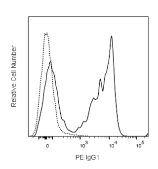
This H-2K[b]:Ig fusion protein has been tested by immunofluorescent staining (≤ 4 µg H-2K[b]:Ig/million cells) (see Figure) and flow cytometric analysis of antigen-specific T cells to assure specificity and reactivity. It is necessary to load the H-2K[b] portions of the dimeric protein with a relevant peptide of interest prior to immunofluorescent staining of T cells. H-2K[b]:Ig complexes are effectively loaded by incubation with excess relevant (specific) or irrelevant (control) peptides (see Protocol 1). Peptide-loaded H-2Kb:Ig may be used for immunofluorescent staining (see Protocol 2). Since applications vary, each investigator must determine dilutions appropriate for individual use.

Protocol 1: Peptide Loading of H-2K[b]:Ig Dimeric Protein

An alloreactive T-cell clone, 2C, is specific for an endogenous peptide, p2Ca. Several related peptides or peptide analogs have been identified. These differ in their MHC restriction and in their affinity for 2C TCR. For H-2Kb, SIY peptide can also be recognized by 2C TCR in the context of H-2K[b]. SIY has a relatively high affinity for H-2K[b] and is suggested as a positive control for staining of 2C cells in this assay. The 2C clone was originally derived by stimulating BALB/c spleen cells with irradiated P815 (H-2Ld) cells.

Several peptide-loading protocols have been described. The method used at BD Biosciences Pharmingen involves passive loading of excess peptide in solution with H-2K[b]:Ig protein. We have found that passive loading works particularly well in the case of high-affinity peptides. For lower-affinity peptides, an increase in the molar ratio of peptide to H-2K[b]:Ig may improve loading, as determined by flow cytometric analysis. It is suggested that for each peptide, parameters such as the dose of H-2K[b]:Ig per million cells, molar ratio of peptide to H-2K[b]:Ig, and peptide loading time be determined empirically by the investigator. While this BD DimerX product contains β2 Microglobulin, for investigators requiring excess recombinant Human β2 Microglobulin, we recommend BD Biosciences Cat. No. 551089.

Peptide preparation and loading:

1. The molecular weight (MW) of a peptide of interest will need to be determined. A peptide's MW can be estimated by multiplying its number (n) of amino acids (AA) by 130 daltons (d) per amino acid:

        MW of peptide (d) = n (AA) x 130 (d/AA)

2. A stock of peptide may be prepared at 20 mg/ml in DMSO. Dilute the peptide solution to 2 mg/ml in sterile DPBS, pH 7.2 for use in the H-2K[b]:Ig loading protocol.

3. Mix H-2K[b]:Ig protein with specific or control peptide at 40, 160, or 640 molar (M) excess. The following calculation, using an 8 amino acid peptide (8mer) as an example, may be used:

        Dp = Molecular Weight of peptide: e.g., 8 amino acids x 130 = 1,040 daltons.

        DK[b] = Molecular Weight of H-2K[b]:Ig = 250,000 daltons.

        R = desired excess molar ratio, e.g., 160.

        Mp = micrograms (µg) peptide of interest.

        MK[b] = micrograms (µg) H-2K[b]:Ig in the reaction. A typical amount of peptide-loaded H-2K[b]:Ig to use for flow cytometry staining is         0.25 to 4 µg/million cells (test).

        Mp = MK[b] x R x Dp = 4 µg x 160 x 1,040 d = 2.66 µg         Therefore, one would add 2.66 µg of peptide and 4 µg of H-2K[b]:Ig

                        DKb                 250,000 d                                    in solution for the optimal peptide loading of H-2Kb:Ig.

4. Mix peptide and H-2K[b]:Ig together in PBS, pH 7.2, incubate at 37°C overnight. The peptide-loaded H-2K[b]:Ig can be stored at 4°C for up to 1 week.

Protocol 2: Immunofluorescent Staining Protocol

1. Prepare peptide-loaded H-2K[b] protein staining cocktail by mixing 0.25- 4 µg of peptide-loaded H-2K[b] protein/test with 0.25 - 4 µg of PE-conjugated A85-1 mAb (anti-mouse IgG1, Cat. No. 550083)/test at a ratio of 1:1 or 1:2 of dimer:A85-1 mAb. Incubate the mixture for 60 minutes at RT, protect from exposure to light.

2. Add 0.25 - 4 µg of purified mouse IgG1 isotype control mAb A111-3 (Cat. No. 553485)/test to the staining cocktail (see Step 1 above). Incubate the staining cocktail for 30 minutes at RT, protect from exposure to light.

3. Resuspend mouse cells in BD FACS™ staining buffer [eg, DPBS, 1% FCS, 0.09% NaN3 or BD Pharmingen™ Stain Buffer (FBS), Cat. No. 554656], containing the appropriate amount of Mouse BD Fc Block™ purified anti-mouse CD16/CD32 mAb 2.4G2 (Cat. No. 553141/553142), at a concentration of approximately 10e6 cells per 50 µl. Incubate 10 minutes at 4°C. Add ~1 x10e6 cells per staining tube (eg, 12 x 75 mm tube, BD Falcon™ Cat. No. 352008).

4. Add 50 µl BD FACS buffer containing the optimal per test amount of the staining cocktail, plus any other cell-surface marker-specific antibodies to be used to each sample.

5. Wash cells 2x with 2 ml BD FACS buffer, centrifuge for 5 minutes at 250 x g, and discard supernatant. Resuspend cell pellet in approximately 0.5 ml staining buffer in a tube appropriate for the flow cytometer.

Protocol 3: Alternative: Immunofluorescent Staining Protocol

1. Resuspend mouse cells in BD FACS staining buffer [e.g., DPBS, 1% FCS, 0.09% NaN3 or BD Pharmingen Stain Buffer (FBS), Cat. No. 554656], containing the appropriate amount of Mouse BD Fc Block purified anti-mouse CD16/CD32 mAb 2.4G2 (Cat. No. 553141/553142), at a concentration of approximately 10e6 cells per 50 µl. Incubate 10 minutes at 4°C. Add ~1 x 10e6 cells per staining tube (eg, 12 x 75 mm tube, BD Falcon Cat. No. 352008).

2. Add 0.25 - 4 µg of peptide-loaded H-2K[b]:Ig protein to cell suspension. Incubate 60 minutes at 4°C.

3. Wash cells 1x with 2 ml BD FACS buffer, centrifuge for 5 minutes at 250 µg, and aspirate supernatant.

4. Resuspend cells in 100 µl BD FACS buffer containing appropriately diluted fluorescent secondary reagent. We typically use PE-conjugated A85-1 mAb (anti-mouse IgG1, Cat. No. 550083). Incubate 30 - 60 minutes at 4°C.

5. Wash cells 2x with 2 ml BD FACS buffer, centrifuge for 5 minutes at 250 x g, and discard supernatant. Resuspend cell pellet in approximately 0.5 ml staining buffer in a tube appropriate for the flow cytometer.

Product Notices

  1. Since applications vary, each investigator should titrate the reagent to obtain optimal results.
  2. Please refer to www.bdbiosciences.com/us/s/resources for technical protocols.
  3. Caution: Sodium azide yields highly toxic hydrazoic acid under acidic conditions. Dilute azide compounds in running water before discarding to avoid accumulation of potentially explosive deposits in plumbing.
550750 Rev. 1
Antibody Details
Down Arrow Up Arrow
DimerX/H-2Kb

The H-2K[b]:Ig fusion protein consists of three extracellular major histocompatibility complex (MHC) class I H-2Kb domains that are fused to the VH regions of mouse IgG1. In order for the MHC class I to be functional, i.e., capable of binding peptides, β2 Microglobulin (β2M) must be present. For this reason, BD™ DimerX consists of recombinant H-2K[b]:Ig fusion protein, supplemented with recombinant β2M. Recombinant MHC molecules, like the BD DimerX fusion protein, are useful for studying T-cell function by immunofluorescent staining and flow cytometric analysis of antigen-specific T cells.

The MHC gene locus encodes a group of highly polymorphic, cell-surface proteins that play a broad role in the immune response to protein antigens. MHC molecules function by binding and presenting small antigenic protein fragments to antigen-specific receptors expressed by T cells (TCR). Human (human leukocyte antigen/HLA) and mouse (histocompatibility 2/H-2) MHC molecules are structurally and functionally related proteins that comprise two major classes. Class I MHC molecules consist of two separate polypeptide chains. The class I α chain is an MHC encoded, transmembrane polypeptide containing three extracellular domains: α1, α2, and α3. The second chain consists of a non-MHC encoded polypeptide called β2M. Since β2M does not contain a transmembrane domain, it associates with the α chain through noncovalent interaction. Functionally, class I MHC molecules can bind peptides derived from intracellular antigens (e.g., viral and some bacterial antigens) that are specifically recognized by CD8+ T cells. Class II MHC molecules consist of two different transmembrane proteins that can bind peptide fragments derived from extracellular proteins (e.g., bacteria and fungi) and are specifically recognized by CD4+ T cells. TCR recognize both processed peptides bound to MHC, as well as regions of the MHC molecule itself. CD4 and CD8 accessory molecules strengthen formation of the TCR-MHC complex through their interaction with nonpolymorphic regions of the MHC molecule.

550750 Rev. 1
Format Details
Down Arrow Up Arrow
Purified
Tissue culture supernatant is purified by either protein A/G or affinity purification methods. Both methods yield antibody in solution that is free of most other soluble proteins, lipids, etc. This format provides pure antibody that is suitable for a number of downstream applications including: secondary labeling for flow cytometry or microscopy, ELISA, Western blot, etc.
Purified
550750 Rev.1
Citations & References
Down Arrow Up Arrow

Development References (4)

  1. Cai Z, Brunmark AB, Luxembourg AT, et al. Probing the activation requirements for naive CD8+ T cells with Drosophila cell transfectants as antigen presenting cells. Immunol Rev. 1998; 165:249-265. (Biology). View Reference
  2. Cai Z, Kishimoto H, Brunmark A, Jackson MR, Peterson PA, Sprent J. Requirements for peptide-induced T cell receptor downregulation on naive CD8+ T cells. J Exp Med. 1997; 185(4):641-651. (Biology). View Reference
  3. Dal Porto J, Johansen TE, Catipovic B, et al. A soluble divalent class I major histocompatibility complex molecule inhibits alloreactive T cells at nanomolar concentrations. Proc Natl Acad Sci U S A. 1993; 90(14):6671-6675. (Biology). View Reference
  4. Schneck JP, Slansky JE, O'Herrin SM, Greten TF . Monitoring antigen-specific T cells using MHC-Ig dimers. In: Coligan J, Kruisbeek D, Margulies EM, Shevach EM, Strober W, ed. Current Protocols in Immunology. New York: John Wiley & Sons, Inc; 2000:17.2.1-17.2.17.
View All (4) View Less
550750 Rev. 1

Please refer to Support Documents for Quality Certificates


Global - Refer to manufacturer's instructions for use and related User Manuals and Technical data sheets before using this products as described


Comparisons, where applicable, are made against older BD Technology, manual methods or are general performance claims.  Comparisons are not made against non-BD technologies, unless otherwise noted.

For Research Use Only. Not for use in diagnostic or therapeutic procedures.

Refer to manufacturer's instructions for use and related User Manuals and Technical Data Sheets before using this product as described.

Comparisons, where applicable, are made against older BD technology, manual methods or are general performance claims. Comparisons are not made against non-BD technologies, unless otherwise noted.