


Product Details
Description
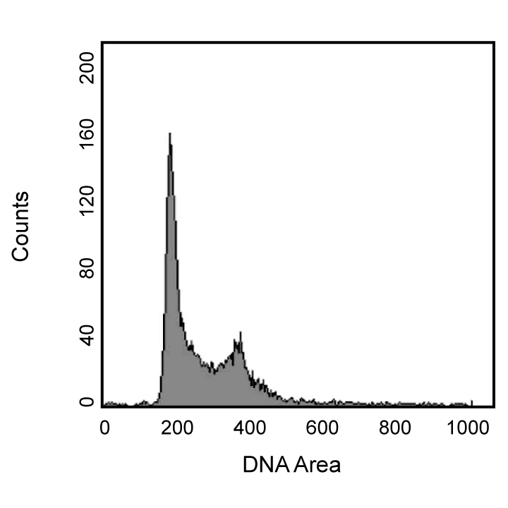
Propidium Iodide (PI) is a fluorescent vital dye that stains DNA and RNA. PI binds to both DNA and RNA, so the latter must be removed by digestion with ribonucleases. The content of DNA as determined by flow cytometry, and can reveal useful information about the cell cycle and the proteins involved in cell cycle regulation. Cells in G2 and M phases of the cell cycle have double the DNA content of those in G0 and G1 phases. Cells in S phase have DNA content lying between these extremes. PI is detected in the orange range of the spectrum using a 562-588 nm band pass filter. This reagent may be used to analyze cell cycle by flow cytometry in addition to use with antibodies for examining the expression of proteins during the cell cycle.
Preparation And Storage
Recommended Assay Procedures
Flow cytometry: After fixing and permeabilizing your cell sample, use 0.5 mL /test (1 x 10e6 cells) and incubate for 15 minutes at room temperature before analysis. Please refer to http://static.bdbiosciences.com/documents/BD_FlowCytometry_DNA_Staining_Protocol.pdf for more protocol information.
Product Notices
- Caution: Sodium azide yields highly toxic hydrazoic acid under acidic conditions. Dilute azide compounds in running water before discarding to avoid accumulation of potentially explosive deposits in plumbing.
- Avoid contact with skin and eyes.
- Please refer to http://regdocs.bd.com to access safety data sheets (SDS).
- Please refer to www.bdbiosciences.com/us/s/resources for technical protocols.
Please refer to Support Documents for Quality Certificates
Global - Refer to manufacturer's instructions for use and related User Manuals and Technical data sheets before using this products as described
Comparisons, where applicable, are made against older BD Technology, manual methods or are general performance claims. Comparisons are not made against non-BD technologies, unless otherwise noted.
For Research Use Only. Not for use in diagnostic or therapeutic procedures.
Refer to manufacturer's instructions for use and related User Manuals and Technical Data Sheets before using this product as described.
Comparisons, where applicable, are made against older BD technology, manual methods or are general performance claims. Comparisons are not made against non-BD technologies, unless otherwise noted.