BD Pharmingen™ Purified Mouse Anti-Lck w/ Control
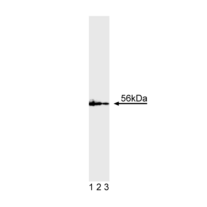
Western blot analysis of lck expression on Jurkat cell lysate. Jurkat cell lysate (Cat. No. 611451) was probed with Purified Mouse Anti-Lck (Cat. No. 51-1495GR) at concentrations of 2.0 (lane 1), 1.0 (lane 2), and 0.5 µg/ml (lane 3). Lck was visualized with HRP Goat Anti-Mouse Ig (Cat. No. 554002), and identified as a band of 56 kDa.
Western blot analysis of lck expression on Jurkat cell lysate. Jurkat cell lysate (Cat. No. 611451) was probed with Purified Mouse Anti-Lck (Cat. No. 51-1495GR) at concentrations of 2.0 (lane 1), 1.0 (lane 2), and 0.5 µg/ml (lane 3). Lck was visualized with HRP Goat Anti-Mouse Ig (Cat. No. 554002), and identified as a band of 56 kDa.
Description
The T-cell receptor (TCR) is a multi-chain transmembrane receptor responsible for antigen recognition on the T-cell surface. T cells also express several other integral membrane proteins, including CD4 and CD8, which play significant roles in the functional responses of the TCR to antigen presentation. Following antigen or ligand binding to the TCR, a series of interrelated membrane and cytoplasmic activation or signaling events rapidly occurs. These events include tyrosine phosphorylation of membrane and cytoplasmic proteins, plasma membrane inositol phospholipid hydrolysis, increases in cytoplasmic calcium concentrations, and increases in protein kinase C activity. The earliest measurable biochemical changes are the appearance of newly phosphorylated tyrosine residues on a variety of cytoplasmic and membrane proteins. Phosphorylation of tyrosine residues is mediated by protein tyrosine kinases (PTKs). Several different PTKs have been implicated in early phosphorylation events of T cell activation, including lck. p56-lck is a member of the Src family, and its functional domains can be divided into two regions based on sequence comparison with p60-src. The N-terminal half, which is highly divergent, contains the membrane bound/substrate interactive domain. The C-terminal half, which has more conserved homology, contains the kinase domain. Lck is normally expressed exclusively in cells of lymphoid lineage, primarily T cells, and natural killer cells. Lck is generally expressed at detectable levels in T-cell lines, including CTLL-2 (ATCC TIB-214) and Jurkat. Lower levels of lck expression have been detected in B cells. Aberrant expression of lck has been described in human colon and lung carcinoma cell lines. Lck plays a role T-cell signal transduction through its physical association with the cytoplasmic tails of CD4 and CD8 expressed in helper and cytolytic T-cells, respectively. The importance of lck in T cell activation is supported by genetic studies. For example, mice with lck null mutations lack T cell development. Additionally, mice expressing only mutant CD4 that is unable to bind to lck, lack the ability to activate T cells through the TCR. MOL 171 reacts with human lck proteins (56-60 kDa). It cross-reacts with mouse lck proteins. A 25 amino acid synthetic peptide corresponding to the N-terminal region of the human lck sequence was used as immunogen.
Preparation And Storage
Recommended Assay Procedures
Applications include western blot analysis (0.5-2.0 µg/ml). Other applications not routinely tested at BD Biosciences Pharmingen include immunoprecipitation (2.0-5.0 µg/ml). Lck is typically detected as one or multiple bands ranging between 56-60 kDa. Jurkat control lysate [50 µg (1 µg/µl)] is provided as a western blot positive control (Cat. No. 51-16526N; store lysate at -20°C). Additional control lysate (Cat. No. 611451) is sold separately. U937 human histiocytic lymphoma cells (ATCC CRL-1593) are suggested as a negative control. For western blot analysis, 5 µg total cell protein per lane is recommended for SDS/PAGE mini-gels. Non-specific bands may be detected if too much protein is loaded onto the gel.
Product Notices
- Since applications vary, each investigator should titrate the reagent to obtain optimal results.
- Caution: Sodium azide yields highly toxic hydrazoic acid under acidic conditions. Dilute azide compounds in running water before discarding to avoid accumulation of potentially explosive deposits in plumbing.
- Source of all serum proteins is from USDA inspected abattoirs located in the United States.
- Species cross-reactivity detected in product development may not have been confirmed on every format and/or application.
- Sodium azide is a reversible inhibitor of oxidative metabolism; therefore, antibody preparations containing this preservative agent must not be used in cell cultures nor injected into animals. Sodium azide may be removed by washing stained cells or plate-bound antibody or dialyzing soluble antibody in sodium azide-free buffer. Since endotoxin may also affect the results of functional studies, we recommend the NA/LE (No Azide/Low Endotoxin) antibody format, if available, for in vitro and in vivo use.
- Please refer to http://regdocs.bd.com to access safety data sheets (SDS).
- Please refer to www.bdbiosciences.com/us/s/resources for technical protocols.
Companion Products

| Description | Quantity/Size | Part Number | EntrezGene ID |
|---|---|---|---|
| N/A | 50.0 | N/A | N/A |
Please refer to Support Documents for Quality Certificates
Global - Refer to manufacturer's instructions for use and related User Manuals and Technical data sheets before using this products as described
Comparisons, where applicable, are made against older BD Technology, manual methods or are general performance claims. Comparisons are not made against non-BD technologies, unless otherwise noted.
For Research Use Only. Not for use in diagnostic or therapeutic procedures.