BD Pharmingen™ Purified NA/LE Mouse Anti-Human Neuropilin-1 (CD304)
Clone mAb3 (also known as MAB3; mAb#3) (RUO)
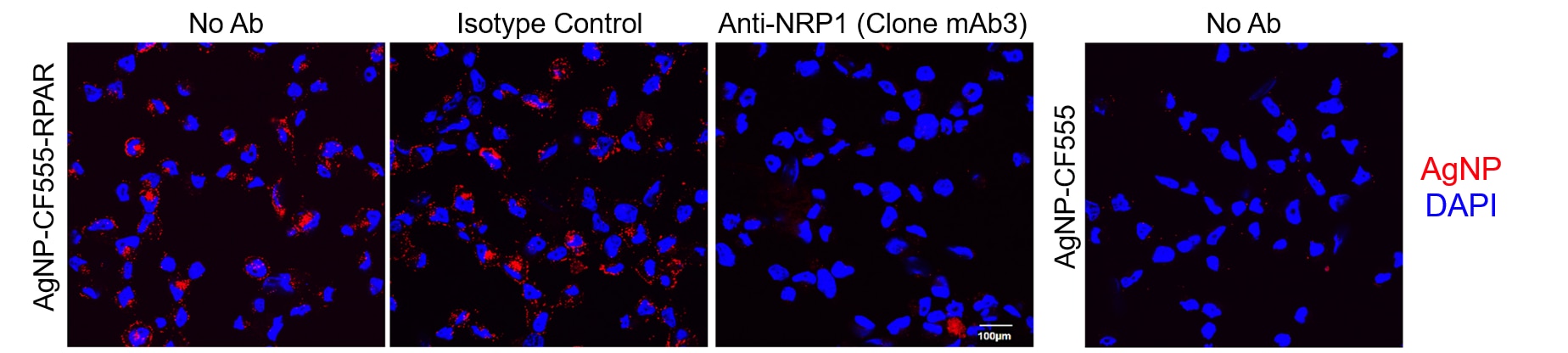
Clone Mab3 blocks the binding of prototypic CendR peptide RPARPAR Nanoparticles to Neuropilin-l (NRPI). Cells from the Human PPC-1I prostate cancer cell line expressing NRP-I were pre-incubated (30 min at 37 °C) with either No Antibody, Purified NA/LE Mouse IgG1, κ Isotype Control (Isotype Control; Cat. No. 554721; 30 µg/ml) or Purified NA/LE Mouse Anti-Human Neuropilin-1 (CD304) [Anti-NRP1 (Clone mAb3); Cat. No. 570225; 30 µg/ml] as indicated. Nanoparticles displaying prototypic CendR peptide RPARPAR (AgNP-CF555-RPAR) were then incubated (60 min; 37 °C) with the cells. After 3 washes with PBS, the cells were fixed (1 min; -20 °C) with methanol. After two more washes with PBS, cells were counterstained with DAPI before imaging by confocal microscopy (60x). Nanoparticles without prototypic CendR peptide RPARPAR (AgNP-CF555) were used as a negative control. Data was kindly provided by Allan Tobi and Tambet Teesalu.
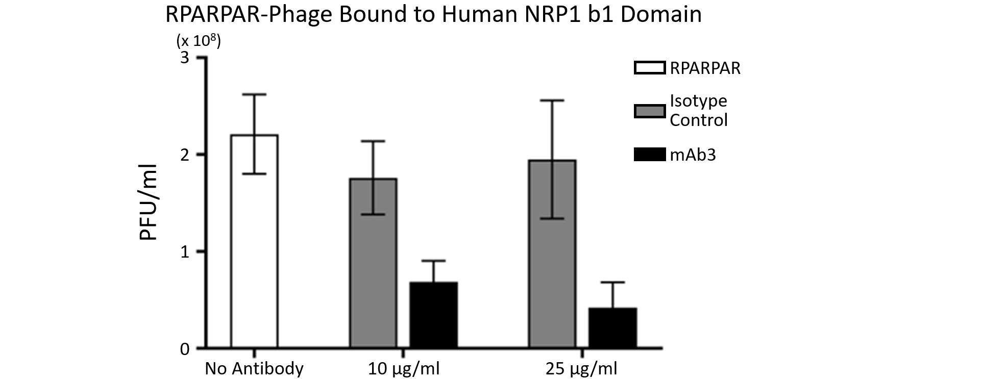
Clone Mab3 blocks the binding of T7 bacteriophages expressing prototypic CendR peptide RPARPAR to hNRP-1 b1b2 domain. Magnetic beads coated with His-tagged Human NRP1 b1 domain were incubated (60 min; Room Temperature) with either No Antibody, Purified NA/LE Mouse IgG1, κ Isotype Control (Isotype Control; Cat. No. 554721), or Purified NA/LE Mouse Anti-Human Neuropilin-1 (CD304) [Clone mAb3; Cat. No. 570225] at 10 µg/ml or 25 µg/ml as indicated. After washing, the beads were incubated (60 min; Room Temperature) with 10^9 T7 bacteriophages displaying prototypic CendR peptide RPARPAR. After further washing, bound T7 bacteriophages were eluted and titered. Data was kindly provided by Jan Lukas Hobrock, Kaarel Kurm and Tambet Teesalu.
Clone Mab3 blocks the binding of prototypic CendR peptide RPARPAR Nanoparticles to Neuropilin-l (NRPI). Cells from the Human PPC-1I prostate cancer cell line expressing NRP-I were pre-incubated (30 min at 37 °C) with either No Antibody, Purified NA/LE Mouse IgG1, κ Isotype Control (Isotype Control; Cat. No. 554721; 30 µg/ml) or Purified NA/LE Mouse Anti-Human Neuropilin-1 (CD304) [Anti-NRP1 (Clone mAb3); Cat. No. 570225; 30 µg/ml] as indicated. Nanoparticles displaying prototypic CendR peptide RPARPAR (AgNP-CF555-RPAR) were then incubated (60 min; 37 °C) with the cells. After 3 washes with PBS, the cells were fixed (1 min; -20 °C) with methanol. After two more washes with PBS, cells were counterstained with DAPI before imaging by confocal microscopy (60x). Nanoparticles without prototypic CendR peptide RPARPAR (AgNP-CF555) were used as a negative control. Data was kindly provided by Allan Tobi and Tambet Teesalu.
Clone Mab3 blocks the binding of T7 bacteriophages expressing prototypic CendR peptide RPARPAR to hNRP-1 b1b2 domain. Magnetic beads coated with His-tagged Human NRP1 b1 domain were incubated (60 min; Room Temperature) with either No Antibody, Purified NA/LE Mouse IgG1, κ Isotype Control (Isotype Control; Cat. No. 554721), or Purified NA/LE Mouse Anti-Human Neuropilin-1 (CD304) [Clone mAb3; Cat. No. 570225] at 10 µg/ml or 25 µg/ml as indicated. After washing, the beads were incubated (60 min; Room Temperature) with 10^9 T7 bacteriophages displaying prototypic CendR peptide RPARPAR. After further washing, bound T7 bacteriophages were eluted and titered. Data was kindly provided by Jan Lukas Hobrock, Kaarel Kurm and Tambet Teesalu.
Preparation And Storage
Product Notices
- Please refer to www.bdbiosciences.com/us/s/resources for technical protocols.
- Since applications vary, each investigator should titrate the reagent to obtain optimal results.
- Please refer to http://regdocs.bd.com to access safety data sheets (SDS).
Companion Products

The mAb3 monoclonal antibody specifically recognizes Neuropilin-1 (NRP1), also known as CD304, Blood dendritic cell antigen 4 (BDCA4), and Vascular endothelial cell growth factor 165 receptor (VEGF165R). Neuropilin-1 (CD304) is an ~140 kDa type I transmembrane glycoprotein that is encoded by NRP1 and expressed on plasmacytoid dendritic cells (DC), thymocytes, regulatory T cells, a subset of T follicular helper cells, endothelial cells, neurons, and certain tumor cells. This multifunctional receptor can mediate the interaction, growth, survival, and migration of a variety of normal and tumor cells including prostate and breast cancer cells. Neuropilin-1 (CD304) may play a role in the interactions between some T cells and dendritic cells. It is involved in the development of the nervous and cardiovascular systems. CD304 complexes with Plexin-A family members to bind chemorepellent Class 3 Semaphorins and guide neuronal axon growth. It also functions as a coreceptor with VEGFR2/CD309 to stimulate angiogenesis in response to VEGF165. It also functions as a co-receptor that can facilitate the entry of severe acute respiratory syndrome coronavirus 2 (SARS-CoV-2) into host cells.
Development References (7)
-
Bruder D et al. Neuropilin-1: a surface marker of regulatory T cells.. Eur J Immunol. 2004; 34(3):623-630. (Biology). View Reference
-
Daly JL, Simonetti B, Klein K, et al. Neuropilin-1 is a host factor for SARS-CoV-2 infection. Science. 2020; 370(6518):861-865. (Immunogen). View Reference
-
Dzionek A et al. BDCA-2, BDCA-3, and BDCA-4: three markers for distinct subsets of dendritic cells in human peripheral blood.. J Immunol. 2000; 165(11):6037-6046. (Biology). View Reference
-
Mizui M, Kumanogoh A, Kikutani H. Immune semaphorins: novel features of neural guidance molecules.. J Clin Immunol. 2009; 29(1):1-11. (Biology). View Reference
-
Pan Q et al. Neuropilin-1 binds to VEGF121 and regulates endothelial cell migration and sprouting.. J Biol Chem. 2007; 282(33):24049-24056. (Biology). View Reference
-
Renand A, Milpied P, Rossignol J, et al. Neuropilin-1 expression characterizes T follicular helper (Tfh) cells activated during B cell differentiation in human secondary lymphoid organs.. PLoS ONE. 2013; 8(12):e85589. (Biology). View Reference
-
Soker S, Takashima S, Miao HQ, Neufeld G, Klagsbrun M. Neuropilin-1 is expressed by endothelial and tumor cells as an isoform-specific receptor for vascular endothelial growth factor.. Cell. 1998; 92(6):735-45. (Biology). View Reference
Please refer to Support Documents for Quality Certificates
Global - Refer to manufacturer's instructions for use and related User Manuals and Technical data sheets before using this products as described
Comparisons, where applicable, are made against older BD Technology, manual methods or are general performance claims. Comparisons are not made against non-BD technologies, unless otherwise noted.
For Research Use Only. Not for use in diagnostic or therapeutic procedures.