

Preparation And Storage
Recommended Assay Procedures
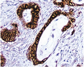
Immunohistochemistry: Clone OX-38 is tested for immunohistochemical staining of acetone-fixed frozen sections of rat spleen or thymus. IHC of formalin-fixed paraffin embedded sections is not recommended. This antibody has been reported to stain the CD4 subset of T lymphocytes. The isotype control recommended for use with Purified Mouse IgG2a κ Isotype Control (Cat. No. 550339). For optimal indirect immunohistochemical staining, the OX-38 antibody should be titrated (1:10 to 1:50 dilution) and visualized via a three-step staining procedure in combination with Biotin Rat Anti-Mouse IgG2a(Cat. No. 550332) as the secondary antibody and Streptavidin-HRP (Cat. No. 550946) together with DAB Substrate Kit (Cat. No. 550880).
Product Notices
- Since applications vary, each investigator should titrate the reagent to obtain optimal results.
- An isotype control should be used at the same concentration as the antibody of interest.
- Source of all serum proteins is from USDA inspected abattoirs located in the United States.
- Caution: Sodium azide yields highly toxic hydrazoic acid under acidic conditions. Dilute azide compounds in running water before discarding to avoid accumulation of potentially explosive deposits in plumbing.
- Sodium azide is a reversible inhibitor of oxidative metabolism; therefore, antibody preparations containing this preservative agent must not be used in cell cultures nor injected into animals. Sodium azide may be removed by washing stained cells or plate-bound antibody or dialyzing soluble antibody in sodium azide-free buffer. Since endotoxin may also affect the results of functional studies, we recommend the NA/LE (No Azide/Low Endotoxin) antibody format, if available, for in vitro and in vivo use.
- This antibody has been developed for the immunohistochemistry application. However, a routine immunohistochemistry test is not performed on every lot. Researchers are encouraged to titrate the reagent for optimal performance.
- Please refer to www.bdbiosciences.com/us/s/resources for technical protocols.
Companion Products


The OX-38 antibody specifically recognizes the CD4 antigen on most thymocytes, a subpopulation of mature T lymphocytes (ie, MHC class II-restricted T cells, including most T helper cells), monocytes, macrophages, and some dendritic cells. CD4 is an antigen coreceptor on the T-cell surface which interacts with MHC class II molecules on antigen-presenting cells. It participates in T-cell activation through its association with the T-cell receptor complex and protein tyrosine kinase lck. The OX-38 antibody has been reported to bind to the same epitope of CD4 as that recognized by W3/25 mAb, which is a different epitope than that recognized by OX-35 mAb. In vivo blocking of some cell-mediated immune responses by mAb OX-38 has been reported. Injection of OX-38 mAb induces allograft unresponsiveness in rats, with varying results depending on the rat strain used (high or low responder). Furthermore, in vivo depletion of CD4+ lymphocytes has been reported with this antibody.
Development References (9)
-
Arima T, Goss JA, Walp LA, Flye MW. Administration of anti-CD4 monoclonal antibody with intrathymic injection of alloantigen results in rat cardiac allograft tolerance. Surgery. 1995; 118(2):265-273. (Clone-specific: Depletion). View Reference
-
Bañuls MP, Alvarez A, Ferrero I, Zapata A, Ardavin C. Cell-surface marker analysis of rat thymic dendritic cells. Immunology. 1993; 79(2):298-304. (Biology). View Reference
-
Bierer BE, Sleckman BP, Ratnofsky SE, Burakoff SJ. The biologic roles of CD2, CD4, and CD8 in T-cell activation. Annu Rev Immunol. 1989; 7:579-599. (Biology). View Reference
-
Janeway CA Jr. The T cell receptor as a multicomponent signalling machine: CD4/CD8 coreceptors and CD45 in T cell activation. Annu Rev Immunol. 1992; 10:645-674. (Biology). View Reference
-
Jefferies WA, Green JR, Williams AF. Authentic T helper CD4 (W3/25) antigen on rat peritoneal macrophages. J Exp Med. 1985; 162(1):117-127. (Immunogen: Flow cytometry, Functional assay, Immunoaffinity chromatography, Immunoprecipitation, Inhibition). View Reference
-
Liu L, Zhang M, Jenkins C, MacPherson GG. Dendritic cell heterogeneity in vivo: two functionally different dendritic cell populations in rat intestinal lymph can be distinguished by CD4 expression. J Immunol. 1998; 161(3):1146-1155. (Biology). View Reference
-
Stitz L, Sobbe M, Bilzer T. Preventive effects of early anti-CD4 or anti-CD8 treatment on Borna disease in rats. J Virol. 1992; 66(6):3316-3323. (Clone-specific: Blocking). View Reference
-
Suzuki H, Hara MH, Miyahara T, et al. Microchimerism and graft acceptance: IV. Cardiac allograft acceptance following anti-adhesion molecule antibody therapy. Transplant Proc. 1996; 28(4):2058-2060. (Clone-specific: Blocking). View Reference
-
Yin D, Fathman CG. Tissue-specific effects of anti-CD4 therapy in induction of allograft unresponsiveness in high and low responder . Transpl Immunol. 1995; 3(3):258-264. (Clone-specific: Blocking). View Reference
Please refer to Support Documents for Quality Certificates
Global - Refer to manufacturer's instructions for use and related User Manuals and Technical data sheets before using this products as described
Comparisons, where applicable, are made against older BD Technology, manual methods or are general performance claims. Comparisons are not made against non-BD technologies, unless otherwise noted.
For Research Use Only. Not for use in diagnostic or therapeutic procedures.