Skip to main content Skip to navigation
Purified Hamster Anti-Mouse CD119
Expression of cell surface IFN-γRα chains by BALB/c splenic lymphocytes. BALB/c spleen cells (including erythrocytes) were preincubated (~15 minutes, 4°C) with purified 2.4G2 antibody [BD Fc Block™; Cat. No. 553141/553142; 1 µg antibody/10e6 cells]. The cells were stained (30 minutes, 4°C) with purified 2E2 antibody (0.5 µg mAb/10e6 cells; Cat. No. 559911). After washing, the cells were incubated (30 minutes, 4°C) with a biotin-conjugated cocktail of mouse anti-hamster antibodies (Clones G70-204 + G94-56; cat. No. 554010; 0.5 µg mAb cocktail/10e6 cells). Finally, the cells were washed and incubated with R-PE-conjugated Streptavidin (Cat. No. 554061; 0.015 µg PE-SA/10e6 cells) and FITC-anti-CD4 (clone RM4-5; Cat. No. 553047) and FITC-anti-CD8 (clone 53-6.7; Cat. No. 553031), both at 0.06 µg mAb/10e6 cells. After washing, the cells were analyzed with a FACScan™ Flow Cytometer. The immunofluorescent staining patterns for erythrocytes and lymphocytes that were either not stained with 2E2 (top images, background staining) or were stained with purified 2E2 (bottom images) followed by the 2nd and 3rd layer reagents shown at right. The two-color dot plots were generated by gating for cells that had the light-scattering characteristics of erythrocytes (left images) or lymphocytes (right images). The data indicates that mouse erythrocytes are uniformly negative for IFN-γRα expression whereas most T cells (i.e., CD4+/CD8+ cells) and CD4-CD8- lymphocytes constitutively express medium levels of IFN-γRα chains.
Product Details
Down Arrow Up Arrow


BD Pharmingen™
Ifngr1; IFN-γ Receptor α; IFN-gamma R alpha; Ifngr; INF-g Receptor; Cd119
Mouse (QC Testing)
Armenian Hamster IgG1, κ
Purified preparation of soluble recombinant mouse IFN-γRα chain protein
Flow cytometry (Routinely Tested), Immunoprecipitation (Reported)
0.5 mg/ml
AB_397366
Aqueous buffered solution containing ≤0.09% sodium azide.
RUO


Preparation And Storage

The monoclonal antibody was purified from tissue culture supernatant or ascites by affinity chromatography. Store undiluted at 4°C.

Recommended Assay Procedures

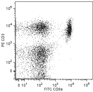
Immunofluorescent Staining and Flow Cytometric Analysis: The purified form of 2E2 (Cat. No. 559911) can be used for the immunofluorescent staining (≤ 1 µg antibody/10e6 cells) and flow cytometric analysis of normal mouse cells or cell lines to measure their expressed levels of IFN-γRα. An appropriate purified immunoglobulin isotype control is A19-3. A three-layer staining protocol is recommended for maximizing the detection IFN-γRα chains expressed by cells as detailed in the figure legend.

Note: 2E2 is a nonblocking antibody that can be used for the unobstructed immunofluorescent staining and flow cytometric analysis of cells in systems where the ligand (i.e., IFN-γ) for IFN-γ receptors is present.

Immunoprecipitation: The 2E2 antibody has been reported to be useful for the immunoprecipitation of IFN-γRα chains from lysates of cloned mouse T cells.

Product Notices

  1. Since applications vary, each investigator should titrate the reagent to obtain optimal results.
  2. Please refer to www.bdbiosciences.com/us/s/resources for technical protocols.
  3. Caution: Sodium azide yields highly toxic hydrazoic acid under acidic conditions. Dilute azide compounds in running water before discarding to avoid accumulation of potentially explosive deposits in plumbing.
559911 Rev. 1
Antibody Details
Down Arrow Up Arrow
2E2

The 2E2 antibody recognizes the extracellular region of the 90 kDa alpha chain subunit of the mouse interferon-γ receptor (IFN-γRα; aka, CD119). The functionally active-form of the mouse IFN-γ receptor consists of two (or more) subunits, with IFN-γRα responsible for IFN-γ binding and both the IFN-γRα and IFN-γRβ chains required for the transduction of biologic responses. IFN-γRα is expressed by a variety of cell lines and normal mouse cells (except mature erythrocytes) including T cells, B cells, NK cells, monocytes, neutrophils, fibroblasts, epithelial and endothelial cells. The 2E2 antibody is a non-neutralizing antibody; it does not block the binding of IFN-γ to its receptor. The immunogen used to generate this hybridoma was a purified preparation of soluble recombinant mouse IFN-γRα chain protein.

559911 Rev. 1
Format Details
Down Arrow Up Arrow
Purified
Tissue culture supernatant is purified by either protein A/G or affinity purification methods. Both methods yield antibody in solution that is free of most other soluble proteins, lipids, etc. This format provides pure antibody that is suitable for a number of downstream applications including: secondary labeling for flow cytometry or microscopy, ELISA, Western blot, etc.
Purified
559911 Rev.1
Citations & References
Down Arrow Up Arrow

Development References (1)

  1. Bach EA, Szabo SJ, Dighe AS, et al. Ligand-induced autoregulation of IFN-gamma receptor beta chain expression in T helper cell subsets.. Science. 1995; 270(5239):1215-8. (Clone-specific: Immunoprecipitation). View Reference
559911 Rev. 1

Please refer to Support Documents for Quality Certificates


Global - Refer to manufacturer's instructions for use and related User Manuals and Technical data sheets before using this products as described


Comparisons, where applicable, are made against older BD Technology, manual methods or are general performance claims.  Comparisons are not made against non-BD technologies, unless otherwise noted.

For Research Use Only. Not for use in diagnostic or therapeutic procedures.