Due to some routine maintenance you might experience temporary service interruptions or slowness on
Sunday, July 19, 2026, 2:00 - 10:00 am (EDT).
If you encounter problems while ordering, please contact us at BDBCustomerService@bd.com.
See More
Due to some routine maintenance you might experience temporary service interruptions or slowness on
Sunday, July 19, 2026, 2:00 - 10:00 am (EDT).
If you encounter problems while ordering, please contact us at BDBCustomerService@bd.com.
See MoreDue to some routine maintenance you might experience temporary service interruptions or slowness on
Sunday, July 19, 2026, 2:00 - 10:00 am (EDT)
.If you encounter problems while ordering, please contact us at BDBCustomerService@bd.com.
Looks like you're visiting us from {countryName}.
Would you like to stay on the current country site or be switched to your country?
Two-color flow cytometric analysis of IFN-γ expression in stimulated human peripheral blood lymphocytes. Human peripheral blood mononuclear cells were stimulated for 6 hours with Phorbol 12-Myristate 13-Acetate (Sigma P-8139; 50 ng/ml final concentration) and Ionomycin (Sigma I-0634; 1 μg/ml final concentration) in the presence of BD GolgiStop™ Protein Transport Inhibitor (containing Monensin) (Cat. No. 554724). The cells were harvested, washed with BD Pharmingen™ Stain Buffer (FBS) (Cat. No. 554656), and fixed with BD Cytofix™ Fixation Buffer (Cat. No. 554655). The cells were then washed and stained in BD Perm/Wash™ Buffer (Cat. No. 554723) with BD Horizon™ BV421 Mouse Anti-Human CD3 antibody (Cat. No. 562426/562427) and either BD Horizon™ BB700 Mouse IgG1, κ Isotype Control (Cat. No. 566404; Left Plot) or BD Horizon BB700 Mouse Anti-Human IFN-γ antibody (Cat. No. 566394/566395; Right Plot). Two-color flow cytometric dot plots showing the correlated expression of IFN-γ (or Ig Isotype control staining) versus CD3 were derived from gated events with the forward and side light-scatter characteristics of intact lymphocytes. Flow cytometric analysis was performed using a BD LSRFortessa™ X-20 Cell Analyzer System.
Any use of products other than the permitted use without the express written authorization of Becton, Dickinson and Company is strictly prohibited.
For optimal and reproducible results, BD Horizon Brilliant Stain Buffer should be used anytime two or more BD Horizon Brilliant dyes are used in the same experiment. Fluorescent dye interactions may cause staining artifacts which may affect data interpretation. The BD Horizon Brilliant Stain Buffer was designed to minimize these interactions. More information can be found in the Technical Data Sheet of the BD Horizon Brilliant Stain Buffer (Cat. No. 563794 or 566349).
When setting up compensation, it is recommended to compare spillover values obtained from cells and BD™ CompBeads to ensure that beads will provide sufficiently accurate spillover values.
For optimal results, it is recommended to perform two washes after staining with antibodies. Cells may be prepared, stained with antibodies and washed twice with wash buffer per established protocols for immunofluorescent staining, prior to acquisition on a flow cytometer. Performing fewer than the recommended wash steps may lead to increased spread of the negative population.
The B27 monoclonal antibody specifically binds to human interferon-γ (IFN-γ), a 14-18 kDa glycoprotein containing 143 amino acid residues. IFN-γ is a potent multifunctional cytokine produced by several activated cell types including NK, NKT, CD4+TCRαβ+, CD8+TCRαβ+, and TCRγδ+ T cells. IFN-γ exerts its biological effects through specific binding to the high-affinity IFN-γ receptor complex comprised of IFN-γRα (CD119) and IFN-γRβ subunits. In addition to its antiviral effects, IFN-γ upregulates a number of lymphoid cell functions including the antimicrobial and anti-tumor responses of macrophages, NK cells, and neutrophils. In addition, IFN-γ influences the regulation of proliferation, differentiation, and effector responses of B cell and T cell subsets. These influences can involve IFN-γ's capacity to boost MHC class I and II expression by antigen-presenting cells as well as direct effects on B cells and T cells themselves. B27 is a neutralizing antibody. The use of B27 antibody for epitope mapping of human IFN-γ has been described. The B27 antibody has been reported not to bind to denatured IFN-γ.
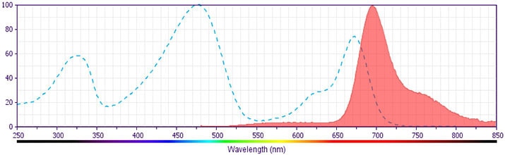
The antibody was conjugated to BD Horizon BB700, which is part of the BD Horizon Brilliant™ Blue family of dyes. It is a polymer-based tandem dye developed exclusively by BD Biosciences. With an excitation max of 485 nm and an emission max of 693 nm, BD Horizon BB700 can be excited by the 488 nm laser and detected in a standard PerCP-Cy™5.5 set (eg, 695/40-nm filter). This dye provides a much brighter alternative to PerCP-Cy5.5 with less cross laser excitation off the 405 nm and 355 nm lasers.
Please refer to Support Documents for Quality Certificates
Global - Refer to manufacturer's instructions for use and related User Manuals and Technical data sheets before using this products as described
Comparisons, where applicable, are made against older BD Technology, manual methods or are general performance claims. Comparisons are not made against non-BD technologies, unless otherwise noted.

For Research Use Only. Not for use in diagnostic or therapeutic procedures.