T-bet (O4-46)
Select Conjugate
About T-bet
ISOTYPE
IgG1, k
CYTOMETER USED
FACSCantoII
EXPERIMENT CELL SOURCE
Human PBMC (Fresh Blood)
REACTIVE SPECIES
Human, Mouse
HOST SPECIES
Mouse
NCBI ENTREZ INFO
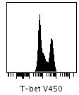
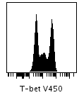
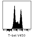
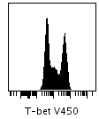
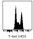
T-bet V450
| Perm I | Perm II | Perm III | Perm IV (0.5x) | Perm IV (1.0x) | |
|---|---|---|---|---|---|
| Lymphocytes |
|
|
|
|
|
Ratio between the MFI of stimulated vs control
| Perm I | Perm II | Perm III | Perm IV (0.5x) | Perm IV (1.0x) | |
|---|---|---|---|---|---|
| Lymphocytes | 2.68 | 3.39 | 3.5 | 3.1 | 3.08 |
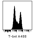
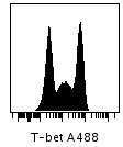
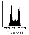
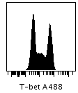
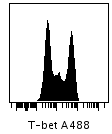
T-bet A488
| Perm I | Perm II | Perm III | Perm IV (0.5x) | Perm IV (1.0x) | |
|---|---|---|---|---|---|
| Lymphocytes |
|
|
|
|
|
Ratio between the MFI of stimulated vs control
| Perm I | Perm II | Perm III | Perm IV (0.5x) | Perm IV (1.0x) | |
|---|---|---|---|---|---|
| Lymphocytes | 3.09 | 3.94 | 4.06 | 3.43 | 3.38 |
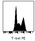
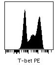
T-bet PE
| Perm I | Perm II | Perm III | Perm IV (0.5x) | Perm IV (1.0x) | |
|---|---|---|---|---|---|
| Lymphocytes |
|
|
|
|
|
Ratio between the MFI of stimulated vs control
| Perm I | Perm II | Perm III | Perm IV (0.5x) | Perm IV (1.0x) | |
|---|---|---|---|---|---|
| Lymphocytes | 3.6 | 4.25 | 4.33 | 3.78 | 3.72 |
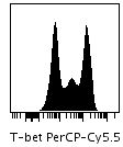
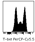
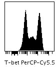
T-bet PerCP-Cy5.5
| Perm I | Perm II | Perm III | Perm IV (0.5x) | Perm IV (1.0x) | |
|---|---|---|---|---|---|
| Lymphocytes |
|

|
|
|
|
Ratio between the MFI of stimulated vs control
| Perm I | Perm II | Perm III | Perm IV (0.5x) | Perm IV (1.0x) | |
|---|---|---|---|---|---|
| Lymphocytes | 4.07 | 3.88 | 3.98 | 3.86 | 3.85 |
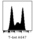
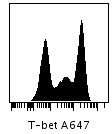
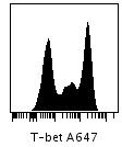
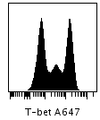
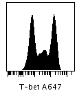
T-bet A647
| Perm I | Perm II | Perm III | Perm IV (0.5x) | Perm IV (1.0x) | |
|---|---|---|---|---|---|
| Lymphocytes |
|
|
|
|
|
Ratio between the MFI of stimulated vs control
| Perm I | Perm II | Perm III | Perm IV (0.5x) | Perm IV (1.0x) | |
|---|---|---|---|---|---|
| Lymphocytes | 4.6 | 4.42 | 4.53 | 4.11 | 4.07 |
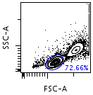
Gating Hierarchy
Lymphocytes
 Lymphocytes
Protocol & Other Details
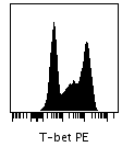
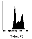
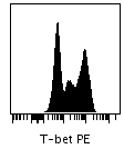
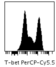
T-bet expression varies among human lymphocyte subsets, with up to three levels of expression being detected in an optimal stain. The T-bet–high population is made up largely of NK cells and effector T cells. Most memory T cells are found in the T-bet–int population, whereas naïve T cells are negative for T-bet expression.
Notes
Human Surface Marker Antibody Concentrations:
Antibody titration is critical for successful post-permeabilization staining of CD markers and other cell surface antigens. The 1X concentration represents cells stained with the recommended test size for live cell stains (see product Technical Data Sheets for test size information). The 1/4X or 1/16X concentrations represent cells stained with 1/4 or 1/16 the antibody recommended for live cell stains.
Mouse Surface Marker Antibody Concentrations:
Antibody titration is critical for successful post-permeabilization staining of CD markers and other cell surface antigens. The 1X concentration represents cells stained with the recommended test size for live cell stains. The 1/4X or 1/16X concentrations represent cells stained with 1/4 or 1/16 the antibody recommended for live cell stains.
YG PE, YG PE-Cy7, and YG PE-Cy5:
PE, PE-Cy5, and PE-Cy7 fluorochromes may be excited by either a 488-nm blue laser or a 561-nm yellow-green laser. Since excitation by the yellow-green laser improves PE detection sensitivity, data collected from the 561-nm laser detection array (YG PE, YG PE-Cy5, and YG PE-Cy7) are provided along with data from the 488-nm laser detection array (PE, PE-Cy5, and PE-Cy7) when available.
Fold-Change Statistic:
Fold-change of a channel (e.g. pStat5-PE) is calculated as the ratio between the median fluorescence intensity of the stimulated sample (e.g. IL-2) and the median fluorescence intensity of the unstimulated sample (e.g. Control).
Percentile Distance Statistic:
Percentile distance is a measure of the spread between the 95th percentile and the 5th percentile of the fluorescence intensity of a channel (e.g. CD3-FITC). The distance is calculated as the ratio between the 95th percentile of a channel and the 5th percentile of a channel in its transformed space (log, linear, arcsinh or biexponential).
Permeabilization Buffers
BD Phosflow™ Perm/Wash Buffer I (557885)
BD Phosflow™ Perm Buffer II (558052)
BD Phosflow™ Perm Buffer III (558050)
BD Phosflow™ Perm Buffer IV (560746)
Perm Buffer IV 1X vs. 0.5X:
Perm Buffer IV (provided as a 10X stock solution) may be used at a 1X or 0.5X concentration. The higher (1X) concentration provides optimal resolution of certain intracellular phosphoprotein stains but might result in increased cell loss and decreased ability to stain certain cell surface markers.
Entrez/About
About this Protein/NCBI Entrez Information:
30009
Keywords:
T-box expressed in T cells, TBX21, T-box 21, TBLYM
For Research Use Only. Not for use in diagnostic or therapeutic procedures.
23-23343-00