Products
-
Instruments
-
Flow Cytometers
- Clinical Cell Analyzers
-
Research Cell Analyzers
- BD® LSR II Flow Cytometer
- BD FACSCelesta™ Cell Analyzer
- BD FACSLyric™ Research System
- LSRFortessa™ Cell Analyzer
- LSRFortessa™ X-20
- FACSymphony™ A5
- BD Accuri™ C6
- FACSVerse™
- FACSymphony™ A3
- BD Accuri™ C6 Plus
- FACSymphony™ A5 SE Cell Analyzer
- FACSymphony™ A1 Cell Analyzer
- BD FACSDiscover™ A8 Research Cell Analyzer
- Research Cell Sorters
- Clinical Sample Prep Systems
- Single-Cell Multiomics Systems
-
Flow Cytometers
-
Reagents
-
Flow Cytometry Reagents
- Clinical Diagnostics
-
Research Reagents
- BD Horizon RealViolet™ 828 for Flow Cytometry
- Quality and Reproducibility
- Single Color Antibodies RUO
- Panels Multicolor Cocktails RUO
- Flow Cytometry Controls and Lysates
- buffers and Supporting Reagents RUO
- Cell Function Analysis Stains Dyes
- Single Color Antibodies
- Compensation Beads
- BD Horizon™ Human T Cell Backbone Panel
- BD Pharmingen™ MonoBlock™ Leukocyte Staining Buffer
- BV605 Transition
- BD Horizon RealBlue™ 670 for Flow Cytometry
- BD Horizon RealBlue™ 780 for Flow Cytometry
- BD Horizon RealYellow™ 586
- BD Horizon RealYellow™ 610
- BD Horizon RealYellow™ 703
- Clinical Discovery
-
Western Blotting and Molecular Reagents
- Immunoassay Reagents
-
Single-Cell Multiomics Reagents
- BD® AbSeq Assay
-
BD® Single-Cell Multiplexing Kit
-
BD Rhapsody™ ATAC-Seq Assays
-
BD Rhapsody™ Whole Transcriptome Analysis (WTA) Amplification Kit
-
BD Rhapsody™ TCR/BCR Next Multiomic Assays
-
BD Rhapsody™ Targeted mRNA Kits
-
BD Rhapsody™ Accessory Kits
-
BD Rhapsody™ TCR/BCR Profiling Assays for Human and Mouse
- BD® OMICS-One Protein Panels
-
Functional Assays
-
Microscopy and Imaging Reagents
-
Cell Preparation and Separation Reagents
-
Flow Cytometry Reagents
Solutions
Discover & Learn
Resources & Tools
Support
-
-
- BD® LSR II Flow Cytometer
- BD FACSCelesta™ Cell Analyzer
- BD FACSLyric™ Research System
- LSRFortessa™ Cell Analyzer
- LSRFortessa™ X-20
- FACSymphony™ A5
- BD Accuri™ C6
- FACSVerse™
- FACSymphony™ A3
- BD Accuri™ C6 Plus
- FACSymphony™ A5 SE Cell Analyzer
- FACSymphony™ A1 Cell Analyzer
- BD FACSDiscover™ A8 Research Cell Analyzer
-
-
-
- BD Horizon RealViolet™ 828 for Flow Cytometry
- Quality and Reproducibility
- Single Color Antibodies RUO
- Panels Multicolor Cocktails RUO
- Flow Cytometry Controls and Lysates
- buffers and Supporting Reagents RUO
- Cell Function Analysis Stains Dyes
- Single Color Antibodies
- Compensation Beads
- BD Horizon™ Human T Cell Backbone Panel
- BD Pharmingen™ MonoBlock™ Leukocyte Staining Buffer
- BV605 Transition
- BD Horizon RealBlue™ 670 for Flow Cytometry
- BD Horizon RealBlue™ 780 for Flow Cytometry
- BD Horizon RealYellow™ 586
- BD Horizon RealYellow™ 610
- BD Horizon RealYellow™ 703
-
-
-
- Brazil (English)
-
Change location/language
You are now leaving the BD Biosciences website. The site you are about to visit is operated by a third party. The link to this site neither makes nor implies any representation or warranty for any products or services offered on a third-party site and is intended only to enable convenient access to the third-party site and for no other purpose. Do you want to continue?
Old Browser
For the best web browsing experience, please use Chrome, Safari or Firefox, minimum versions 77.0.3865, 12.1.2 and 68, respectively.
Please confirm your location
Looks like you're visiting us from {countryName}.
Would you like to stay on the current location site or be switched to your location?
Intracellular Staining
Intracellular Staining of Human Red Blood Cells
- For each sample to be stained, fix 10 µl of whole blood in 1 ml cold 0.05% glutaraldehyde for 10 minutes at room temperature. Vortex after addition of cells.
- Wash once with 5 ml staining buffer (PBS with 1%FBS, 0.09%NaN 3). Centrifuge at 200 x g for 5 minutes.
- Resuspend the cell pellet by vortexing in 0.5 ml 0.1% Triton X-100. Incubate for 10 minutes at room temperature.
- Pellet by centrifugation at 200 x g for 5 minutes and aspirate the supernatant.
- Resuspend the red blood cell pellet in 0.5 ml staining buffer.
- Into new tubes, add primary antibody to appropriate concentration in staining buffer to make 100 µl total volume. For example, if the antibody test size volume is 20 µl, add this amount to 80 µl of staining buffer. Add 10 µl of red blood cell suspension to each tube. Incubate for 20 minutes at room temperature (in the dark).
- Wash with 2 ml of staining buffer at 200 x g for 5 minutes.
- For purified and biotin conjugated antibodies: add 50 µl of diluted secondary reagent, incubate at RT for 15-20 minutes. For direct conjugates proceed to step 10.
- Repeat step 7.
- Add 0.5 ml of staining buffer to each tube.
- Proceed to flow cytometric analysis.
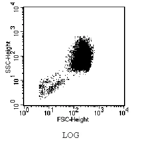
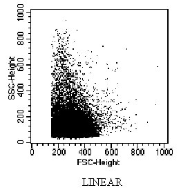
Data Acquisition Note: Setting FSC and SSC detectors to log mode provides better resolution compared to linear mode.