Preparation And Storage
Recommended Assay Procedures
BD® CompBeads can be used as surrogates to assess fluorescence spillover (Compensation). When fluorochrome conjugated antibodies are bound to BD® CompBeads, they have spectral properties very similar to cells. However, for some fluorochromes there can be small differences in spectral emissions compared to cells, resulting in spillover values that differ when compared to biological controls. It is strongly recommended that when using a reagent for the first time, users compare the spillover on cells and BD CompBeads to ensure that BD® CompBeads are appropriate for your specific cellular application.
Product Notices
- Please refer to www.bdbiosciences.com/us/s/resources for technical protocols.
- Caution: Sodium azide yields highly toxic hydrazoic acid under acidic conditions. Dilute azide compounds in running water before discarding to avoid accumulation of potentially explosive deposits in plumbing.
- For fluorochrome spectra and suitable instrument settings, please refer to our Multicolor Flow Cytometry web page at www.bdbiosciences.com/colors.
- Please refer to http://regdocs.bd.com to access safety data sheets (SDS).
- Source of all serum proteins is from USDA inspected abattoirs located in the United States.
- This reagent has been pre-diluted for use at the recommended Volume per Test. We typically use 1 × 10^6 cells in a 100-µl experimental sample (a test).
- An isotype control should be used at the same concentration as the antibody of interest.
Companion Products


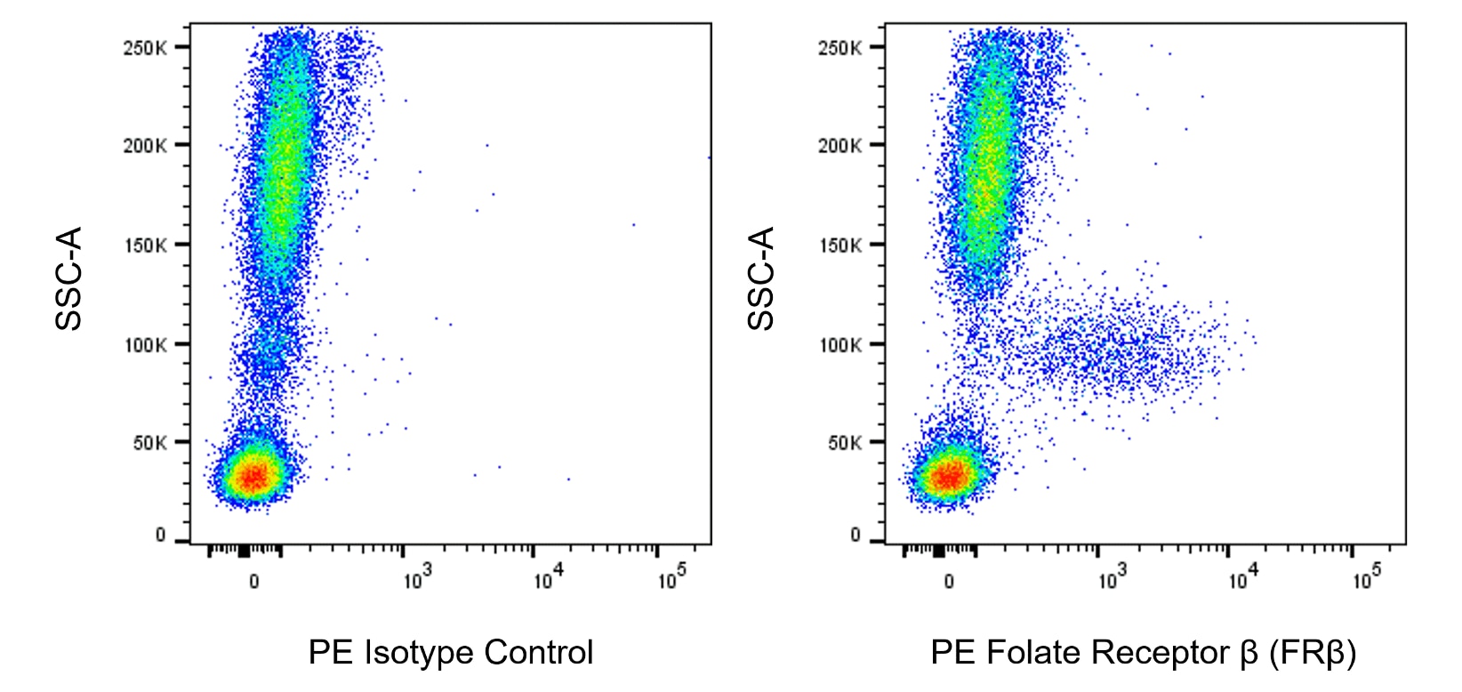
The 94b monoclonal antibody specifically recognizes the human Folate Receptor beta (Folate Receptor β or FRβ). This receptor is a glycosylphosphatidylinositol (GPI)-linked membrane protein that is encoded by FOLR2 which belongs to the folate-binding receptor family. These cell surface receptors share ~70% amino acid sequence homology and bind folate with high affinity. The Folate Receptor β (FRβ) transports folate into cells which is essential for methionine and DNA synthesis that support cell growth and proliferation. FRβ is expressed on placental cells and on monocytes as well as macrophages found in normal and diseased tissues, eg, in rheumatoid arthritis and tumors. Soluble forms of FRβ have also been described which can be found in various body fluids such as plasma or milk.

Development References (5)
-
Holm J, Hansen SI. Characterization of soluble folate receptors (folate binding proteins) in humans. Biological roles and clinical potentials in infection and malignancy.. Biochim Biophys Acta Proteins Proteom. 2020; 1868(10):140466. (Biology). View Reference
-
McNulty H, Pentieva K, Hoey L, Strain J, Ward M. Nutrition throughout life: folate.. Int J Vitam Nutr Res. 2012; 82(5):348-54. (Biology). View Reference
-
Nagai T, Furusho Y, Li H, et al. Production of a High-affinity Monoclonal Antibody Reactive with Folate Receptors Alpha and Beta.. Monoclon Antib Immunodiagn Immunother. 2015; 34(3):181-90. (Clone-specific: Flow cytometry). View Reference
-
Nagayoshi R, Nagai T, Matsushita K, et al. Effectiveness of anti-folate receptor beta antibody conjugated with truncated Pseudomonas exotoxin in the targeting of rheumatoid arthritis synovial macrophages. Arthritis Rheum. 2005; 9(9):2666-2675. (Immunogen). View Reference
-
Otsubo H, Tsuneyoshi Y, Nakamura T, Matsuda T, Komiya S, Matsuyama T. Serum-soluble folate receptor β as a biomarker for the activity of rheumatoid arthritis synovitis and the response to anti-TNF agents.. Clin Rheumatol. 2018; 37(11):2939-2945. (Clone-specific). View Reference
Please refer to Support Documents for Quality Certificates
Global - Refer to manufacturer's instructions for use and related User Manuals and Technical data sheets before using this products as described
Comparisons, where applicable, are made against older BD Technology, manual methods or are general performance claims. Comparisons are not made against non-BD technologies, unless otherwise noted.
For Research Use Only. Not for use in diagnostic or therapeutic procedures.
Refer to manufacturer's instructions for use and related User Manuals and Technical Data Sheets before using this product as described.
Comparisons, where applicable, are made against older BD technology, manual methods or are general performance claims. Comparisons are not made against non-BD technologies, unless otherwise noted.