
Preparation And Storage
Recommended Assay Procedures
BD® CompBeads can be used as surrogates to assess fluorescence spillover (compensation). When fluorochrome conjugated antibodies are bound to BD® CompBeads, they have spectral properties very similar to cells. However, for some fluorochromes there can be small differences in spectral emissions compared to cells, resulting in spillover values that differ when compared to biological controls. It is strongly recommended that when using a reagent for the first time, users compare the spillover on cell and BD® CompBeads to ensure that BD® CompBeads are appropriate for your specific cellular application.
Product Notices
- This reagent has been pre-diluted for use at the recommended Volume per Test. We typically use 1 × 10^6 cells in a 100-µl experimental sample (a test).
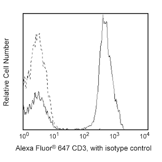
- An isotype control should be used at the same concentration as the antibody of interest.
- Source of all serum proteins is from USDA inspected abattoirs located in the United States.
- Caution: Sodium azide yields highly toxic hydrazoic acid under acidic conditions. Dilute azide compounds in running water before discarding to avoid accumulation of potentially explosive deposits in plumbing.
- For fluorochrome spectra and suitable instrument settings, please refer to our Multicolor Flow Cytometry web page at www.bdbiosciences.com/colors.
- Please refer to http://regdocs.bd.com to access safety data sheets (SDS).
- Please refer to www.bdbiosciences.com/us/s/resources for technical protocols.
Companion Products


The 11A8 monoclonal antibody specifically recognizes DNAX accessory molecule-1 (DNAM-1) which is also known as CD226, Platelet and T cell activation antigen 1 (PTA1), or T lineage-specific activation antigen 1 antigen (TLiSA1). CD226 is an ~65 kDa type I transmembrane glycoprotein that is encoded by CD226 (CD226 antigen) which belongs to the immunoglobulin superfamily (IgSF). The N-terminal extracellular region of CD226 (DNAM-1) contains two C2 type Ig-like domains which are followed by a transmembrane region and a cytoplasmic tail with predicted phosphorylation sites involved in signaling. This adhesion molecule is expressed on subsets of thymocytes, peripheral CD4+ and CD8+ T cells and B cells, NK cells, monocytes, macrophages, and platelets. CD226 (DNAM-1) associates with leukocyte function associated antigen-1 (LFA-1) and is involved in TCR-mediated signal transduction for T cell proliferation and differentiation. Interactions between the CD226 (DNAM-1) activating receptor and its ligands, CD112 and CD155, results in cellular signaling that promotes innate and adaptive immune responses, including the activation, differentiation, and survival of cytotoxic cells.

Development References (3)
-
Cella M, Presti R, Vermi W, et al. Loss of DNAM-1 contributes to CD8+ T-cell exhaustion in chronic HIV-1 infection.. Eur J Immunol. 2010; 40(4):949-54. (Clone-specific: Flow cytometry, Functional assay). View Reference
-
Fourcade J, Sun Z, Chauvin JM, et al. CD226 opposes TIGIT to disrupt Tregs in melanoma.. JCI Insight. 2018; 3(14):121157. (Clone-specific: Flow cytometry). View Reference
-
Fuchs A, Cella M, Giurisato E, Shaw AS, Colonna M. Cutting edge: CD96 (tactile) promotes NK cell-target cell adhesion by interacting with the poliovirus receptor (CD155).. J Immunol. 2004; 172(7):3994-8. (Immunogen: Blocking, Functional assay, Stimulation). View Reference
Please refer to Support Documents for Quality Certificates
Global - Refer to manufacturer's instructions for use and related User Manuals and Technical data sheets before using this products as described
Comparisons, where applicable, are made against older BD Technology, manual methods or are general performance claims. Comparisons are not made against non-BD technologies, unless otherwise noted.
For Research Use Only. Not for use in diagnostic or therapeutic procedures.
Refer to manufacturer's instructions for use and related User Manuals and Technical Data Sheets before using this product as described.
Comparisons, where applicable, are made against older BD technology, manual methods or are general performance claims. Comparisons are not made against non-BD technologies, unless otherwise noted.