-
Reagents
- Flow Cytometry Reagents
-
Western Blotting and Molecular Reagents
- Immunoassay Reagents
-
Single-Cell Multiomics Reagents
- BD® OMICS-Guard Sample Preservation Buffer
- BD® AbSeq Assay
- BD® Single-Cell Multiplexing Kit
- BD Rhapsody™ ATAC-Seq Assays
- BD Rhapsody™ Whole Transcriptome Analysis (WTA) Amplification Kit
- BD Rhapsody™ TCR/BCR Next Multiomic Assays
- BD Rhapsody™ Targeted mRNA Kits
- BD Rhapsody™ Accessory Kits
- BD® OMICS-One Protein Panels
-
Functional Assays
-
Microscopy and Imaging Reagents
-
Cell Preparation and Separation Reagents
-
Dehydrated Culture Media
-
- BD® OMICS-Guard Sample Preservation Buffer
- BD® AbSeq Assay
- BD® Single-Cell Multiplexing Kit
- BD Rhapsody™ ATAC-Seq Assays
- BD Rhapsody™ Whole Transcriptome Analysis (WTA) Amplification Kit
- BD Rhapsody™ TCR/BCR Next Multiomic Assays
- BD Rhapsody™ Targeted mRNA Kits
- BD Rhapsody™ Accessory Kits
- BD® OMICS-One Protein Panels
- Canada (English)
-
Change country/language
Old Browser
Looks like you're visiting us from United States.
Would you like to stay on the current country site or be switched to your country?
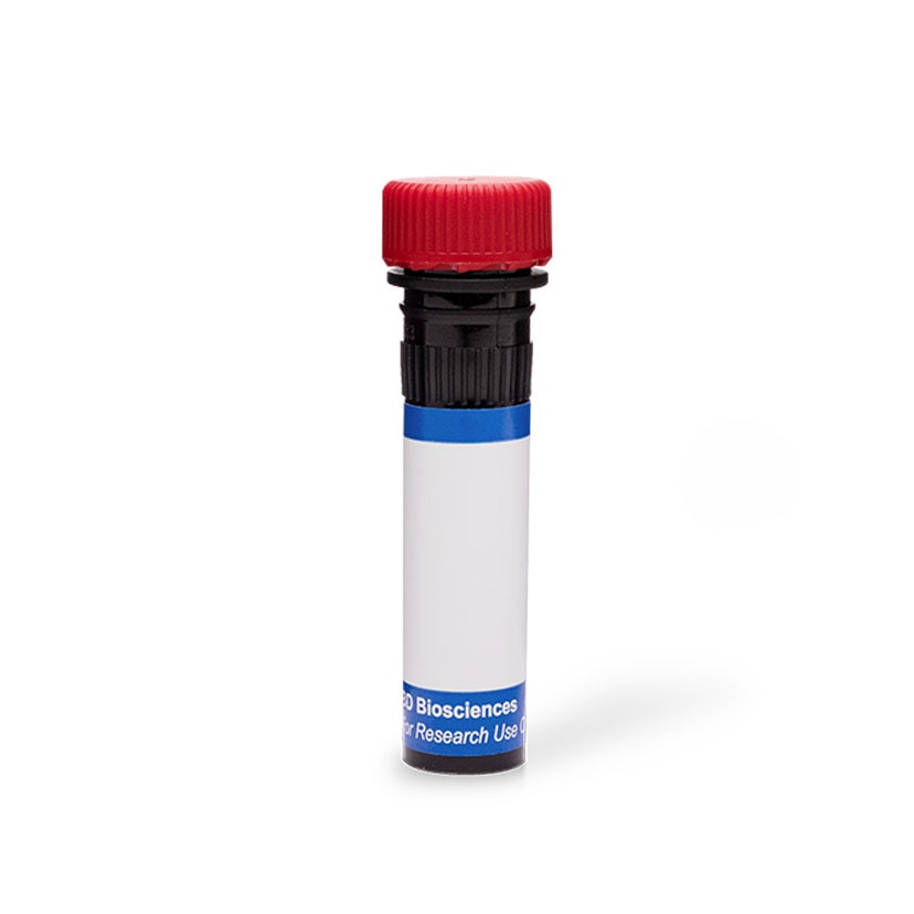
BD Pharmingen™ PE Mouse Anti-Mouse CD289 (TLR9)
Clone J15A7 (RUO)

Multicolor flow cytometric analysis of TLR9 (CD289) expression in mouse splenocytes. Mouse splenic leucocytes were stained with Fixable Viability Stain 450 (FVS450; Cat. No. 562247), fixed, and permeabilized using the BD Cytofix/Cytoperm™ Fixation/Permeabilization Solution Kit (Cat. No. 554714). The cells were then stained with FITC Rat Anti-Mouse CD45R/B220 (Cat. No. 553087/553088/561877) and APC Hamster Anti-Mouse CD11c (Cat. No. 550261/561119) antibodies, and either PE Mouse IgG1, κ Isotype Control (Cat. No. 554680; dotted line histograms) or PE Mouse Anti-Mouse TLR9 antibody (Cat. No. 565640; solid line histograms). The fluorescence histograms (Bottom Plots) showing total cellular TLR9 (CD289) expression [or Ig Isotype control staining] for the different leucocyte subsets were derived from CD11c versus CD45R/B220 coexpression-gated events with the forward and side light-scatter (SSC-A) characteristics of intact FVS450 live cell-discriminated leucocyte subsets (Top Plots). Flow cytometric analysis was performed using a BD LSRFortessa™ Cell Analyzer System.


Multicolor flow cytometric analysis of TLR9 (CD289) expression in mouse splenocytes. Mouse splenic leucocytes were stained with Fixable Viability Stain 450 (FVS450; Cat. No. 562247), fixed, and permeabilized using the BD Cytofix/Cytoperm™ Fixation/Permeabilization Solution Kit (Cat. No. 554714). The cells were then stained with FITC Rat Anti-Mouse CD45R/B220 (Cat. No. 553087/553088/561877) and APC Hamster Anti-Mouse CD11c (Cat. No. 550261/561119) antibodies, and either PE Mouse IgG1, κ Isotype Control (Cat. No. 554680; dotted line histograms) or PE Mouse Anti-Mouse TLR9 antibody (Cat. No. 565640; solid line histograms). The fluorescence histograms (Bottom Plots) showing total cellular TLR9 (CD289) expression [or Ig Isotype control staining] for the different leucocyte subsets were derived from CD11c versus CD45R/B220 coexpression-gated events with the forward and side light-scatter (SSC-A) characteristics of intact FVS450 live cell-discriminated leucocyte subsets (Top Plots). Flow cytometric analysis was performed using a BD LSRFortessa™ Cell Analyzer System.

Multicolor flow cytometric analysis of TLR9 (CD289) expression in mouse splenocytes. Mouse splenic leucocytes were stained with Fixable Viability Stain 450 (FVS450; Cat. No. 562247), fixed, and permeabilized using the BD Cytofix/Cytoperm™ Fixation/Permeabilization Solution Kit (Cat. No. 554714). The cells were then stained with FITC Rat Anti-Mouse CD45R/B220 (Cat. No. 553087/553088/561877) and APC Hamster Anti-Mouse CD11c (Cat. No. 550261/561119) antibodies, and either PE Mouse IgG1, κ Isotype Control (Cat. No. 554680; dotted line histograms) or PE Mouse Anti-Mouse TLR9 antibody (Cat. No. 565640; solid line histograms). The fluorescence histograms (Bottom Plots) showing total cellular TLR9 (CD289) expression [or Ig Isotype control staining] for the different leucocyte subsets were derived from CD11c versus CD45R/B220 coexpression-gated events with the forward and side light-scatter (SSC-A) characteristics of intact FVS450 live cell-discriminated leucocyte subsets (Top Plots). Flow cytometric analysis was performed using a BD LSRFortessa™ Cell Analyzer System.


ImageTitle~BD Pharmingen™ PE Mouse Anti-Mouse CD289 (TLR9)

Regulatory Status Legend
Any use of products other than the permitted use without the express written authorization of Becton, Dickinson and Company is strictly prohibited.
Preparation And Storage
Product Notices
- Since applications vary, each investigator should titrate the reagent to obtain optimal results.
- Please refer to www.bdbiosciences.com/us/s/resources for technical protocols.
- Caution: Sodium azide yields highly toxic hydrazoic acid under acidic conditions. Dilute azide compounds in running water before discarding to avoid accumulation of potentially explosive deposits in plumbing.
- For fluorochrome spectra and suitable instrument settings, please refer to our Multicolor Flow Cytometry web page at www.bdbiosciences.com/colors.
- An isotype control should be used at the same concentration as the antibody of interest.
Data Sheets
Companion Products


The J15A7 monoclonal antibody specifically binds to the mouse Toll-like receptor 9 (TLR9), which is also known as CD289. It recognizes the TLR9N fragment of mouse TLR9 and does not crossreact with human TLR9. TLR9 is a type I transmembrane protein that belongs to the TLR family whose members play fundamental roles in pathogen recognition and activation of innate immunity. TLR9 recognizes unmethylated CpG sequences in DNA molecules. It is expressed by numerous cells of the immune system including B lymphocytes, monocytes, natural killer cells, and subsets of dendritic cells (DC). It is primarily expressed intracellularly within the endosomal compartments. TLR9 functions to alert the immune system of viral and bacterial infections by binding to microbial DNA that is rich in CpG motifs. TLR9 signaling leads to the activation of cells that produce proinflammatory cytokines, such as type-I interferon and IL-12. It can also help induce B cell proliferative responses. TLR9 can reportedly be detected on the surfaces of some DC subsets.

Development References (2)
-
Hemmi H, Takeuchi O, Kawai T, et al. A Toll-like receptor recognizes bacterial DNA. Nature. 2000; 408(6813):740-745. (Biology). View Reference
-
Onji M, Kanno A, Saitoh S, et al. An essential rolw for the N-terminal fragment of Toll-like receptor 9 in DNA sensing. Nat Commun. 2013; 4:1949. (Immunogen: Flow cytometry, Fluorescence microscopy, Immunofluorescence, Immunoprecipitation). View Reference
Please refer to Support Documents for Quality Certificates
Global - Refer to manufacturer's instructions for use and related User Manuals and Technical data sheets before using this products as described
Comparisons, where applicable, are made against older BD Technology, manual methods or are general performance claims. Comparisons are not made against non-BD technologies, unless otherwise noted.
For Research Use Only. Not for use in diagnostic or therapeutic procedures.