





.png)


Preparation And Storage


Recommended Assay Procedures
The 29E.2A3 monoclonal antibody can reportedly bind to Alexa Fluor™ 700 antibody conjugates during multicolor immunofluorescence staining experiments. This interaction may be circumvented by sequentially staining with the 29E.2A3 antibody first, followed by a wash step, and then further staining with an Alexa Fluor™ 700 antibody conjugate of interest.
BD® CompBeads can be used as surrogates to assess fluorescence spillover (Compensation). When fluorochrome conjugated antibodies are bound to BD® CompBeads, they have spectral properties very similar to cells. However, for some fluorochromes there can be small differences in spectral emissions compared to cells, resulting in spillover values that differ when compared to biological controls. It is strongly recommended that when using a reagent for the first time, users compare the spillover on cells and BD® CompBeads to ensure that BD® CompBeads are appropriate for your specific cellular application.
Product Notices
- Please refer to www.bdbiosciences.com/us/s/resources for technical protocols.
- Caution: Sodium azide yields highly toxic hydrazoic acid under acidic conditions. Dilute azide compounds in running water before discarding to avoid accumulation of potentially explosive deposits in plumbing.
- For fluorochrome spectra and suitable instrument settings, please refer to our Multicolor Flow Cytometry web page at www.bdbiosciences.com/colors.
- Please refer to http://regdocs.bd.com to access safety data sheets (SDS).
- This reagent has been pre-diluted for use at the recommended Volume per Test. We typically use 1 × 10^6 cells in a 100-µl experimental sample (a test).
- An isotype control should be used at the same concentration as the antibody of interest.
- Source of all serum proteins is from USDA inspected abattoirs located in the United States.
Companion Products


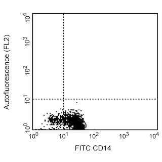
The 29E.2A3 monoclonal antibody specifically recognizes Programmed cell death 1 ligand 1 (PDCD1 ligand 1, PDCD1L1, PDCD1LG1) which is also known as Programmed death ligand 1 (PD-L1 or PDL1) as well as CD274, or B7 homolog 1 (B7-H1, B7H1). PD-L1 (CD274) and PD-L2 (CD273) are type I transmembrane glycoproteins that belong to the B7 family within the Ig gene superfamily and serve as ligands for CD279 (Program Death 1/PD-1). PD-L1 (CD274) is expressed on antigen-presenting cells including activated monocytes, macrophages, and dendritic cells (DCs) as well as activated T cells, B cells, NK cells, and keratinocytes. PD-L1 (CD274) is also variably expressed on placental trophoblasts, myocardial endothelium, cortical thymic epithelial cells, and tumor cells. PD-L1-mediated signaling through PD-1 regulates T cell responses important for providing protective immunity while maintaining peripheral tolerance. This immune signaling checkpoint may also suppress antitumor immune responses and prevent tumor rejection. The 29E.2A3 antibody reportedly blocks PD-L1 (CD274) binding to CD279 (PD-1) and can enhance the proliferation and cytokine production of activated T cells.

Development References (7)
-
Brown JA, Dorfman DM, Ma FR, et al. Blockade of programmed death-1 ligand on dendritic cells enhances T cell activation and cytokine production. J Immunol. 2003; 170:1257-1266. (Clone-specific: Flow cytometry, Functional assay, Immunohistochemistry). View Reference
-
Burr ML, Sparbier CE, Chan YC, et al. CMTM6 maintains the expression of PD-L1 and regulates anti-tumour immunity.. Nature. 2017; 549(7670):101-105. (Clone-specific: Flow cytometry). View Reference
-
Dorfman DM, Brown JA, Shahsafaei A, Freeman GJ. Programmed death-1 (PD-1) is a marker of germinal center-associated T cells and angioimmunoblastic T-cell lymphoma. Am J Surg Pathol. 2006; 30:802-810. (Clone-specific: Immunohistochemistry). View Reference
-
Hughes MJ, McGettrick HM, Sapey E. Importance of validating antibody panels: Anti-PD-L1 clone binds AF700 fluorophore. J Immunol Methods. 2020; 483:112795. (Clone-specific: Flow cytometry). View Reference
-
Latchman Y, Wood CR, Chernova T, et al. PD-L2 is a second ligand for PD-1 and inhibits T cell activation. Nat Immunol. 2001; 2(3):261-268. (Immunogen: ELISA, Flow cytometry). View Reference
-
Llinas L, Lazaro A, de Salort J, Matesanz-Isabel J, Sintes J, Engel P. Expression profiles of novel cell surface molecules on B-cell subsets and plasma cells as analyzed by flow cytometry. Immunol Lett. 2011; 134(2):113-121. (Clone-specific: Flow cytometry). View Reference
-
Rodig N, Ryan T, Allen JA, et al. Endothelial expression of PD-L1 and PD-L2 down-regulates CD8+ T cell activation and cytolysis.. Eur J Immunol. 2003; 33(11):3117-26. (Clone-specific: Flow cytometry, Functional assay). View Reference
Please refer to Support Documents for Quality Certificates
Global - Refer to manufacturer's instructions for use and related User Manuals and Technical data sheets before using this products as described
Comparisons, where applicable, are made against older BD Technology, manual methods or are general performance claims. Comparisons are not made against non-BD technologies, unless otherwise noted.
For Research Use Only. Not for use in diagnostic or therapeutic procedures.
