
Preparation And Storage
Recommended Assay Procedures
It is recommended that cell samples be stained and maintained in the cold (4-8°C) protected from light for flow cytometric analysis.
Product Notices
- This reagent has been pre-diluted for use at the recommended Volume per Test. We typically use 1 × 10^6 cells in a 100-µl experimental sample (a test).
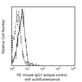
- An isotype control should be used at the same concentration as the antibody of interest.
- For fluorochrome spectra and suitable instrument settings, please refer to our Multicolor Flow Cytometry web page at www.bdbiosciences.com/colors.
- Caution: Sodium azide yields highly toxic hydrazoic acid under acidic conditions. Dilute azide compounds in running water before discarding to avoid accumulation of potentially explosive deposits in plumbing.
- Source of all serum proteins is from USDA inspected abattoirs located in the United States.
- Please refer to www.bdbiosciences.com/us/s/resources for technical protocols.
Companion Products


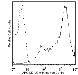
The 17G10.2 monoclonal antibody specifically binds to human Immunoglobulin Like Transcript 7 (ILT7), also known as CD85g. The ILT7 protein is encoded by the LILRA4 (Leukocyte Immunoglobulin Like Receptor subfamily A member 4) gene. ILT7 is a member of immunoglobulin-like transcripts (ILT) or leukocyte immunoglobulin-like receptor (LIR) gene family. ILT7 is selectively expressed on plasmacytoid dendrtic cells (pDC) but not on myeloid dendritic cells or other peripheral blood leukocytes. In response to IL-3, IL-3 receptor complex signaling downregulates ILT7 expression by pDC. ILT7 associates with the FcεRIγ signal adapter protein to form a receptor complex that can signal through activation of an immunoreceptor-based tyrosine activation motif (ITAM)-mediated signaling pathway. Signaling through the ILT7-FcεRIγ receptor complex negatively regulates the innate immune functions of pDC. ILT7 crosslinking inhibits TLR responses by pDC such as the stimulated production of type I interferon and other cytokines. Bone marrow stromal cell antigen 2 (BST2) has been identified as a biological ligand for ILT7.

Development References (3)
-
Cao W, Bover L, Cho M, et al. Regulation of TLR7/9 responses in plasmacytoid dendritic cells by BST2 and ILT7 receptor interaction.. J Exp Med. 2009; 206(7):1603-14. (Biology). View Reference
-
Cao W, Bover L. Signaling and ligand interaction of ILT7: receptor-mediated regulatory mechanisms for plasmacytoid dendritic cells. Immunol Rev. 2010; 234(1):163-176. (Biology). View Reference
-
Cao W, Rosen DB, Ito T, et al. Plasmacytoid dendritic cell-specific receptor ILT7-Fc epsilonRI gamma inhibits Toll-like receptor-induced interferon production.. J Exp Med. 2006; 203(6):1399-405. (Immunogen: Bioassay, Flow cytometry, Inhibition). View Reference
Please refer to Support Documents for Quality Certificates
Global - Refer to manufacturer's instructions for use and related User Manuals and Technical data sheets before using this products as described
Comparisons, where applicable, are made against older BD Technology, manual methods or are general performance claims. Comparisons are not made against non-BD technologies, unless otherwise noted.
For Research Use Only. Not for use in diagnostic or therapeutic procedures.