
Preparation And Storage
Recommended Assay Procedures
Immunofluorescent Staining and Flow Cytometry: The PE Mouse Anti-Human IL-8 (Cat. No. 554720) can be used for multicolor immunofluorescent staining and flow cytometric analyses to identify and enumerate IL-8 producing cells within mixed cell populations (see figure). For optimal immunofluorescent staining with flow cytometric analysis, this anti-cytokine antibody should be titrated (≤ 0.5 µg mAb/million cells). For specific methodology, please visit our protocols under "Intracellular Flow" at our website: http://www.bdbiosciences.com/us/s/resources/.
A useful control for demonstrating specificity of staining is either of the following: 1) pre-block the conjugated G265-8 antibody with ligand (e.g., recombinant human IL-8; Cat. No. 554609) prior to staining, or 2) pre-block the fixed/ permeabilized cells with Purified Mouse Anti-Human IL-8 (Cat. No. 554717/550419) prior to staining. The staining technique and use of blocking controls are described in detail by C. Prussin and D. Metcalfe. An appropriate PE-mouse IgG2b isotype control to use on fixed and permeabilized cells is PE Mouse IgG2b, κ Isotype Control (Cat. No. 555058).
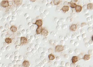
Cytokine ICC: The G265-8 antibody is useful for immunocytochemical staining. Purified Mouse Anti-Human IL-8 (Cat. No. 550419/554717) is tested in the ICC application.
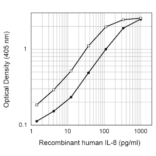
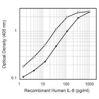
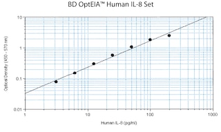
ELISA Detection: The Biotin Mouse Anti-Human IL-8 antibody (Catalog No. 554718) is useful as a detection antibody in a sandwich ELISA for measuring human IL-8 protein levels. Biotin Mouse Anti-Human IL-8 antibody can be paired with the Purified Mouse Anti-Human IL-8 (Cat. No. 554716) with recombinant human IL-8 (Cat. No. 554609) as the standard. This ELISA pair is recommended primarily for measuring cytokine from experimental cell culture systems. For detection of IL-8 in serum or plasma, the Human IL-8 BD OptEIA™ ELISA Set (Cat. No. 555244) or OptEIA™ ELISA Kit (Cat. No. 550999) is recommended.
Product Notices
- Since applications vary, each investigator should titrate the reagent to obtain optimal results.
- An isotype control should be used at the same concentration as the antibody of interest.
- Caution: Sodium azide yields highly toxic hydrazoic acid under acidic conditions. Dilute azide compounds in running water before discarding to avoid accumulation of potentially explosive deposits in plumbing.
- For fluorochrome spectra and suitable instrument settings, please refer to our Multicolor Flow Cytometry web page at www.bdbiosciences.com/colors.
- Please refer to www.bdbiosciences.com/us/s/resources for technical protocols.
Companion Products

The G265-8 monoclonal antibody specifically binds to both the 72 and 77 amino acid isoforms of human Interleukin-8 (IL-8). IL-8 is secreted as an 8-9 kDa, non-glycosylated proinflammatory chemokine protein also known as chemokine (C-X-C motif) ligand 8 (CXCL8). IL-8 is synthesized as a 99 amino acid precursor that is proteolytically processed into several isoforms. The 72 amino acid isoform is produced by monocytes, macrophages, granulocytes, epithelial cells, and fibroblasts in response to pro-inflammatory stimuli including cytokines and microbial agents. It is also expressed by endothelial cells, fibroblasts, keratinocytes, lymphocytes, and a variety of tumor cells. In response to IL-4, IL-10 and TGFβ, the cellular production of IL-8 is inhibited. IL-8 is crucial for the activation and recruitment of neutrophils to inflammatory sites. IL-8 is also a chemoattractant for basophils and T-lymphocytes. IL-8 possesses angiogenic activity and can be associated with tumor angiogenesis and metastasis. The 77 amino acid IL-8 isoform is primarily produced by endothelial cells. This larger isoform is reportedly a less potent neutrophil activator than the 72 amino acid isoform. IL-8 binds to and signals through two G-protein-coupled receptors, IL-8RA (CXCR1/CD181) and IL-8RB (CXCR2/CD182).

Development References (2)
-
Matsushima K, Oppenheim JJ. Interleukin 8 and MCAF: novel inflammatory cytokines inducible by IL 1 and TNF. Cytokine. 1989; 1(1):2-13. (Biology). View Reference
-
Prussin C, Metcalfe DD. Detection of intracytoplasmic cytokine using flow cytometry and directly conjugated anti-cytokine antibodies. J Immunol Methods. 1995; 188(1):117-128. (Methodology). View Reference
Please refer to Support Documents for Quality Certificates
Global - Refer to manufacturer's instructions for use and related User Manuals and Technical data sheets before using this products as described
Comparisons, where applicable, are made against older BD Technology, manual methods or are general performance claims. Comparisons are not made against non-BD technologies, unless otherwise noted.
For Research Use Only. Not for use in diagnostic or therapeutic procedures.