Due to some routine maintenance you might experience temporary service interruptions or slowness on
Sunday, July 19, 2026, 2:00 - 10:00 am (EDT).
If you encounter problems while ordering, please contact us at BDBCustomerService@bd.com.
See More
Due to some routine maintenance you might experience temporary service interruptions or slowness on
Sunday, July 19, 2026, 2:00 - 10:00 am (EDT).
If you encounter problems while ordering, please contact us at BDBCustomerService@bd.com.
See MoreDue to some routine maintenance you might experience temporary service interruptions or slowness on
Sunday, July 19, 2026, 2:00 - 10:00 am (EDT)
.If you encounter problems while ordering, please contact us at BDBCustomerService@bd.com.
Looks like you're visiting us from {countryName}.
Would you like to stay on the current country site or be switched to your country?
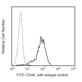
Flow cytometric analysis of CD44 expression on human peripheral blood lymphocytes. Human whole blood was stained with either FITC Mouse Anti-Human CD44 antibody (Cat. No. 555478/560977; solid line histogram) or FITC Mouse IgG2b, κ Isotype Control (Cat. No. 555742; dashed line histogram). The erythrocytes were lysed with BD Pharm Lyse™ Lysing Buffer (Cat. No. 555899). The fluorescence histograms were derived from events with the forward and side light-scatter characteristics of viable lymphocytes. Flow cytometric analysis was performed on a BD FACScan™ (BDIS, San Jose, CA).
Any use of products other than the permitted use without the express written authorization of Becton, Dickinson and Company is strictly prohibited.
The G44-26 monoclonal antibody specifically binds to the 80-95 kDa glycosylated type I transmembrane protein, CD44, also known as phagocytic glycoprotein-1 (Pgp-1). CD44 is the receptor for hyaluronic acid. CD44 is expressed on leucocytes, erythrocytes, epithelial cells and weakly on platelets. CD44 is also called extracellular matrix receptor type III and has functional roles in cell migration, lymphocyte homing and adhesion during hematopoiesis and lymphocyte activation. This antibody recognizes epitope 1 of CD44 antigen according to the HLDA workshop studies.
Please refer to Support Documents for Quality Certificates
Global - Refer to manufacturer's instructions for use and related User Manuals and Technical data sheets before using this products as described
Comparisons, where applicable, are made against older BD Technology, manual methods or are general performance claims. Comparisons are not made against non-BD technologies, unless otherwise noted.
For Research Use Only. Not for use in diagnostic or therapeutic procedures.