







Preparation And Storage


Product Notices
- This reagent has been pre-diluted for use at the recommended Volume per Test. We typically use 1 × 10^6 cells in a 100-µl experimental sample (a test).
- Please refer to www.bdbiosciences.com/us/s/resources for technical protocols.
- Alexa Fluor® is a registered trademark of Molecular Probes, Inc., Eugene, OR.
- Caution: Sodium azide yields highly toxic hydrazoic acid under acidic conditions. Dilute azide compounds in running water before discarding to avoid accumulation of potentially explosive deposits in plumbing.
- For fluorochrome spectra and suitable instrument settings, please refer to our Multicolor Flow Cytometry web page at www.bdbiosciences.com/colors.
- An isotype control should be used at the same concentration as the antibody of interest.
- Source of all serum proteins is from USDA inspected abattoirs located in the United States.
Companion Products




The 7D3 monoclonal antibody specifically binds to T cell immunoglobulin mucin 3 (TIM-3) which is also known as, CD366, or T-cell immunoglobulin and mucin domain-containing protein 3 (TIMD-3/TIMD3). CD366 is encoded by the HAVCR2 gene (Hepatitis A virus cellular receptor 2). CD366 is a type I transmembrane glycoprotein and belongs to the human TIM family (along with TIM-1 and TIM-4) within the immunoglobulin superfamily. CD366 is expressed on Th1, Tc1, Th17, Treg, NK T, and NK cells. CD366 is also expressed on dendritic cells, mast cells, monocytes, and macrophages. It is not expressed by Th2 and B cells. CD366 helps maintain peripheral immune tolerance and homeostasis. CD366 regulates macrophage activation and is a negative regulator of Th1 cell function. Crosslinking of cell surface CD366 by binding to Galectin-9 and/or phosphatidylserine appears to play an important role in either positively or negatively regulating leucocyte functions, such as cytokine production or the phagocytosis of apoptotic cells. CD366 may also be useful as an AML stem cell surface marker because it appears to be more highly expressed by AML leukemia stem cells than by normal bone marrow hematopoietic stem cells.
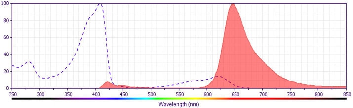
The antibody was conjugated to BD Horizon BV650 which is part of the BD Horizon Brilliant™ Violet family of dyes. This dye is a tandem fluorochrome of BD Horizon BV421 with an Ex Max of 405-nm and an acceptor dye with an Em Max at 650-nm. BD Horizon BV650 can be excited by the violet laser and detected in a filter used to detect APC-like dyes (eg, 660/20-nm filter). Due to the excitation and emission characteristics of the acceptor dye, there will be spillover into the APC and Alexa Fluor® 700 detectors. However, the spillover can be corrected through compensation as with any other dye combination.
Development References (12)
-
Domenig C, Zheng XX, Sabatos CA, et al. Tim-3 inhibits T helper type 1-mediated auto- and alloimmune responses and promotes immunological tolerance. Nat Immunol. 2003; 4(11):1093-1101. (Biology). View Reference
-
Freeman GJ, Casasnovas JM, Umetsu DT, DeKruyff RH. TIM genes: a family of cell surface phosphatidylserine receptors that regulate innate and adaptive immunity.. Immunol Rev. 2010; 235(1):172-89. (Biology). View Reference
-
Hafler DA, Kuchroo V. TIMs: Central regulators of immune responses. J Exp Med. 2008; 205:2699-2701. (Biology). View Reference
-
Jan M, Chao MP, Cha AC, et al. Prospective separation of normal and leukemic stem cells based on differential expression of TIM3, a human acute myeloid leukemia stem cell marker. Proc Natl Acad Sci U S A. 2011; 108(12):5009-5014. (Biology). View Reference
-
Khademi M, Illes Z, Gielen AW, et al. T Cell Ig- and mucin-domain-containing molecule-3 (TIM-3) and TIM-1 molecules are differentially expressed on human Th1 and Th2 cells and in cerebrospinal fluid-derived mononuclear cells in multiple sclerosis. J Immunol. 2004; 172(11):7169-7176. (Biology). View Reference
-
Lee J, Su EW, Zhu C, et al. Phosphotyrosine-dependent coupling of Tim-3 to T-cell receptor signaling pathways. Mol Cell Biol. 2011; 31(19):3963-3974. (Biology). View Reference
-
Lee JS, Park MJ, Park S, Lee ES. Differential expression of T cell immunoglobulin- and mucin-domain-containing molecule-3 (TIM-3) according to activity of Behcet's disease. Br J Dermatol. 2012; 65(3):220-222. (Biology). View Reference
-
Moorman JP, Wang JM, Zhang Y, et al. Tim-3 pathway controls regulatory and effector T cell balance during hepatitis C virus infection. J Immunol. 2012; 189(2):755-766. (Biology). View Reference
-
Ndhlovu LC, Lopez-Verges S, Barbour JD, et al. Tim-3 marks human natural killer cell maturation and suppresses cell-mediated cytotoxicity. Blood. 2012; 119(16):3734-3743. (Biology). View Reference
-
Rodriguez-Manzanet R, DeKruyff R, Kuchroo VK, Umetsu DT. The costimulatory role of TIM molecules. Immunol Rev. 2009; 229(1):259-270. (Biology). View Reference
-
Wang F, Wan L, Zhang C, Zheng X, Li J, Chen ZK. Tim-3-Galectin-9 pathway involves the suppression induced by CD4+CD25+ regulatory T cells. Immunobiology. 2009; 214(5):342-349. (Biology). View Reference
-
van de Weyer PS, Muehlfeit M, Klose C, Bonventre JV, Walz G, Kuehn EW. A highly conserved tyrosine of Tim-3 is phosphorylated upon stimulation by its ligand galectin-9. Biochem Biophys Res Commun. 2006; 351(2):571-576. (Biology). View Reference
Please refer to Support Documents for Quality Certificates
Global - Refer to manufacturer's instructions for use and related User Manuals and Technical data sheets before using this products as described
Comparisons, where applicable, are made against older BD Technology, manual methods or are general performance claims. Comparisons are not made against non-BD technologies, unless otherwise noted.
For Research Use Only. Not for use in diagnostic or therapeutic procedures.
