





.png)


Preparation And Storage


Product Notices
- Since applications vary, each investigator should titrate the reagent to obtain optimal results.
- An isotype control should be used at the same concentration as the antibody of interest.
- Caution: Sodium azide yields highly toxic hydrazoic acid under acidic conditions. Dilute azide compounds in running water before discarding to avoid accumulation of potentially explosive deposits in plumbing.
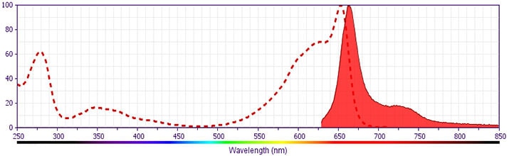
- For fluorochrome spectra and suitable instrument settings, please refer to our Multicolor Flow Cytometry web page at www.bdbiosciences.com/colors.
- Please refer to www.bdbiosciences.com/us/s/resources for technical protocols.
Companion Products




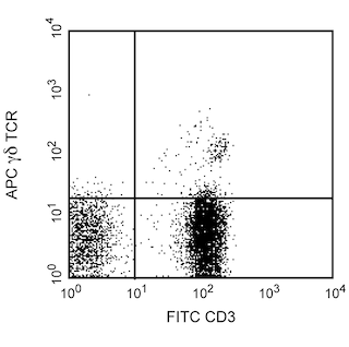
The B1 monoclonal antibody specifically binds to the γδ T cell receptor (γδ TCR). This receptor complex consists of two disulfide-linked transmembrane glycoproteins, a γ chain (45-60 kDa) and a δ subunit (40-60 kDa). The γδ TCR is associated with the signal-transducing CD3 complex. The γδ TCR is expressed by thymocytes and by peripheral T cell subsets (γδ T cells) that are located in the blood, liver, skin and various lymphoid and mucosal tissues. γδ T cells contribute to both innate and adaptive immune responses to infections and tumors. Reports suggest that γδ T cells may also play roles in antigen presentation and the regulation of autoimmune responses.
Development References (10)
-
Bai L, Picard D, Anderson B, et al. The majority of CD1d-sulfatide-specific T cells in human blood use a semiinvariant Vδ1 TCR.. Eur J Immunol. 2012; 42(9):2505-10. (Clone-specific). View Reference
-
Barclay NA, Brown MH, Birkeland ML, et al, ed. The Leukocyte Antigen FactsBook. San Diego, CA: Academic Press; 1997.
-
Bonneville M, O'Brien RL, Born WK. Gammadelta T cell effector functions: a blend of innate programming and acquired plasticity. Nat Rev Immunol. 2110; 10(7):467-478. (Biology). View Reference
-
Breit TM, Wolvers-Tettero IL, van Dongen JJ. Receptor diversity of human T-cell receptor gamma delta expressing cells. Prog Histochem Cytochem. 1992; 26(1-4):182-193. (Biology). View Reference
-
Davodeau F, Peyrat MA, Houde I, et al. Surface expression of two distinct functional antigen receptors on human gamma delta T cells. Science. 1993; 260(5115):1800-1802. (Biology). View Reference
-
De Libero G. Sentinel function of broadly reactive human gamma delta T cells. Immunol Today. 1997; 18(1):22-26. (Biology). View Reference
-
Fitzgerald KA, Callard RE. The cytokine factsbook., 2nd ed. / Katherine A. Fitzgerald ... [et al.].. San Diego: Academic Press; 2001:1-515.
-
Kabelitz D, Pechhold K, Bender A, et al. Activation and activation-driven death of human gamma/delta T cells. Immunol Rev. 1991; 120:71-88. (Biology). View Reference
-
Kabelitz D. Function and specificity of human gamma/delta-positive T cells. Crit Rev Immunol. 1992; 11(5):281-303. (Biology). View Reference
-
Kabelitz D. γδ T-cells: cross-talk between innate and adaptive immunity. Cell Mol Life Sci. 2011; 68(14):2331-2333. (Biology). View Reference
Please refer to Support Documents for Quality Certificates
Global - Refer to manufacturer's instructions for use and related User Manuals and Technical data sheets before using this products as described
Comparisons, where applicable, are made against older BD Technology, manual methods or are general performance claims. Comparisons are not made against non-BD technologies, unless otherwise noted.
For Research Use Only. Not for use in diagnostic or therapeutic procedures.
