
Description
The BD Lyoplate™ Human Cell Surface Marker Screening Panel contains 242 purified monoclonal antibodies to cell surface markers. The panel also contains both mouse and rat isotype controls for assessing isotype-specific background. The panel can be used for screening cell lines, primary cells or tissue, and is compatible with flow cytometry and bioimaging technology platforms. The panel contains three (3) 96 well plates, each well containing 2.75 µg of antibody, enough for five tests (0.5 µg/test) along with AlexaFluor
Component 51-9006585AK - Human Cell Surface Marker Panel - Part A
·Human Cell Surface Marker Lyoplate Plate 1 (1 each) ·Human Cell Surface Marker Lyoplate Plate 2 (1 each) ·Human Cell Surface Marker Lyoplate Plate 3 (1 each) ·Store unopened plates at room temperature (18-25°C). ·Antibodies are lyophilized in an aqueous buffered solution containing BSA and ≤ 0.09% sodium azide.Component 51-9006588BK - Human Cell Surface Marker Screening Panel - Part B
·AlexaFluor® 647 Goat Anti-Mouse Ig (1.8 ml) ·AlexaFluor® 647 Goat Anti-Rat Ig (0.6 ml) ·Store the secondary antibodies at 4°C. ·The secondary antibodies are provided in an aqueous buffered solution containing ≤ 0.09% sodium azide.It is important to note the antibodies present in this panel may not recognize all isoforms of each cell surface marker. In addition, antibody clones can behave differently on cell types depending on the availability of epitopes present, i.e., certain epitopes can be occluded by post-translational modifications. Results you obtain in this screen may only be relevant to the antibody clones tested.
Recommended Assay Procedures
Important instructions before you begin:
Reconstituting the antibody:
·After removing BD Lyoplate Human Cell Surface Marker Screening Panel plates from foil bags, centrifuge at 300 X g for 5 minutes. ·Hold the plate firmly on the work bench and gently remove the foil seal starting from one end and pulling across the plate to completely remove the seal. Once the foil seal is removed, all lyophilized antibodies must be immediately reconstituted. Do not replace the lid on the plate prior to reconstitution. ·Using a multi-channel pipette, reconstitute lyophilized antibodies in 110 µl of 1X sterile PBS. This results in an antibody solution that contains five tests (20 µl/test). Be sure to use fresh pipette tips for each row to prevent well-to-well contamination. Allow antibodies to reconstitute for five minutes at room temperature. ·Store the reconstituted antibodies at 4°C until the cells are prepared for experiments. Reconstituted antibodies can be stored in plates with lids at 4°C for at least 10 days. Seal the plate edges (with lid on) with Parafilm "M"® laboratory film to prevent loss of reconstituted antibody due to evaporation.Screening cells by flow cytometry:
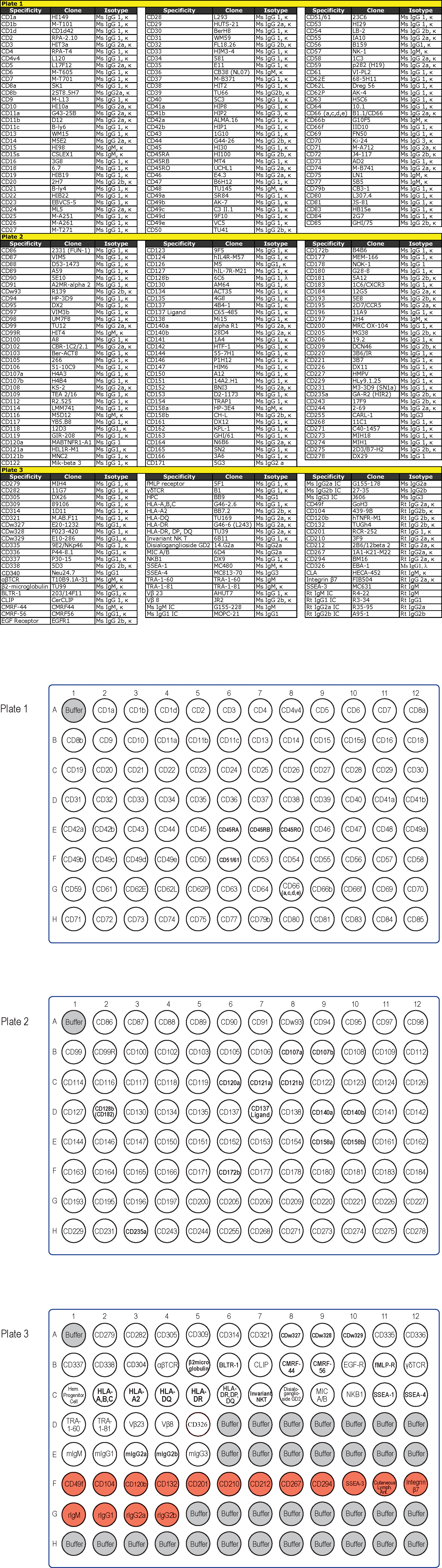
1.Prepare a single cell suspension of live cells from a cell line, tissue or a three dimensional culture. For adherent cell lines, we recommend using either a mild enzyme such as Accutase™ or a non-enzymatic dissociation buffer. 2.Wash the cells in two to four volumes of 1X PBS. Centrifuge at 300 X g for 5 minutes. 3.Remove any clumps by passing the cells through a BD Falcon™ 40 or 70 µm cell strainer (Cat. No. 352340, Cat. No. 352350). 4.Determine the cell concentration and total number of cells. If you are dissociating tissue or a three dimensional culture, we recommend treating the single cells with DNase to prevent cell clumping. Resuspend cells in the recommended growth media or 1X PBS with calcium and magnesium with the addition of 100 units/ml DNase at 10 million cells per ml. Incubate for 15 minutes at room temperature. 5.Wash the cells in two to four volumes of 1X PBS. Centrifuge at 300 X g for 5 minutes. 6.Prepare 275 ml of BD Pharmingen Stain Buffer (FBS) with the addition of 5 mM EDTA (final concentration) for subsequent steps. 7.Resuspend the sample in BD Pharmingen Stain Buffer + EDTA. You will need 135 to 270 million cells (in approximately 27 ml total volume) to fill the antibody containing wells of the three plates (500,000-1,00,000 cells per well). The minimum number of cells per well will depend on the cytometer and/or loss of cells during washing. We have been successful in running as few as 250,000 cells per well. 8.Label three BD Falcon™ round bottom 96 well plates (Cat No. 353910) plates 1, 2 and 3 for your sample plates. 9.Using a multi-channel pipette aliquot 100 µl of cell solution to required wells of the three labeled round-bottom 96-well plates. a.If you have a limited number of cells, you can omit buffer only wells from plate 3. Please refer to the Plate 3 map to identify wells that can be excluded taking into consideration unstained cells and secondary antibody controls. 10.Using a multi-channel pipette, pipette up and down 2-3 times to fully mix the reconstituted antibody from the first row of wells from the BD Lyoplate Screening Panel Plate 1. Add 20 µl to the cells in the corresponding wells of sample plate 1. Continue to add reconstituted antibody to the corresponding sample wells for all remaining wells of each plate. Use fresh tips for every well. Incubate on ice for 20-30 minutes 11.To wash, add 100 µl of BD Pharmingen Stain Buffer + EDTA to each well. Centrifuge at 300 X g for 5 minutes. 12.Remove supernatant carefully and wash cells with an additional 200 µl of BD Pharmingen Stain Buffer + EDTA. Centrifuge at 300 X g for 5 minutes. 13.During the centrifugation step of the final wash, dilute the secondary antibody 1:200 dilution (1.25 µg/ml) in BD Pharmingen Stain Buffer + EDTA. You will need about 26 ml of dilute anti-mouse secondary antibody and about 3 ml of diluted anti-rat secondary antibody. 14.Remove supernatant and apply 100 µl of the appropriate secondary antibody directly to cells in each well and incubate for 20-30 minutes on ice in the dark. a.Add anti-mouse secondary antibody to all wells of the first two labeled round-bottom 96-well sample plates. For sample plate 3, please refer to the Plate maps and add anti-mouse secondary antibody to the appropriate sample wells (white wells: all wells in rows A, B and C wells D1-D5 and E1-E5) and add anti-rat secondary antibody to the appropriate wells (red wells: wells F1-F12 and G1-G4). b.Use remaining wells in sample plate 3 that do not contain antibody (gray wells) to set up unstained cells and anti-rat secondary antibody controls. 15.To wash, add 100 µl of BD Pharmingen Stain Buffer + EDTA to each well. Centrifuge at 300 X g for 5 minutes. 16.Remove supernatant and wash cells with an additional 200µl of BD Pharmingen Stain Buffer + EDTA. Centrifuge at 300 x g for 5 minutes. 17.At this point you may wish to fix your cells prior to analysis. To fix, remove supernatant and add 100 µl of 4% paraformaldehyde in 1X PBS or BD Cytofix™ Fixation Buffer (Cat. No. 554655) per well and incubate for 10 minutes. If you do not wish to fix your cells go to step 19. 18.Wash cells twice with 1X PBS. Centrifuge at 300 X g for 5 minutes. 19.Remove supernatant and resuspend cells in 150 µl of BD Pharmingen Stain Buffer + EDTA per well. Analyze your samples on a flow cytometer. We recommend collecting at least 10,000 events per well. While the first plate is being read, store the other plates on ice in the dark. 20.Analysis templates are available at bdbiosciences.com/resources/stemcell under Tools.Screening cells by bioimaging:
1.Seed the cells in appropriate culture medium at an appropriate cell density in a BD Falcon™ 96-well Imaging Plate (Cat. No. 353219), and culture cells to an appropriate density. We recommend 70-80% confluency for imaging screens. 2.BD Lyoplate surface staining should be performed on live cells as fixation can cause artifacts (false positive and negative signals) with some cell surface markers. In cases where cells must be fixed prior to staining, we recommend confirming any positive hits with a live sample stain using imaging or flow cytometry. 3.Using a multi-channel pipette add 20 µl of each reconstituted antibody to the correspnding wells of your sample plates and incubate on ice for 20-30 minutes. Stain cells directly in 50 to 100 µl of fresh growth media. If staining fixed cells, stain cells in 1X PBS. 4.Wash cells twice in 100 µl 1X PBS. 5.Dilute secondary antibodies to 1:100 (2.5 µg/mL) in growth media and apply 100 µl of the appropriate secondary antibody directly to cells in each well of the sample plates and incubate for 20-30 minutes on ice in the dark. You will need about 26 ml of diluted anti-mouse secondary antibody and about 3 ml of diluted anti-rat secondary antibody. a.Add anti-mouse secondary antibody to all wells of the first two labeled imaging 96-well sample plates. For sample plate 3, please refer to the Plate maps and add anti-mouse secondary antibody to the appropriate sample wells (white wells: all wells in rows A, B and C wells D1-D5 and E1-E5) and add anti-rat secondary antibody to the appropriate wells (red wells: wells F1-F12 and G1-G4). b.Use remaining wells in sample plate 3 that do not contain antibody (grey wells) to set up unstained cells and anti-rat secondary antibody controls. 6.Remove supernatant and wash cells twice in 100 µl 1X PBS. 7.At this point you may wish to fix your cells prior to analysis. To fix, remove supernatant and add 100 µl of 4% paraformaldehyde in 1X PBS or BD Cytofix Fixation Buffer per well and incubate for 10 minutes. If you do not wish to fix your cells go to step 9. 8.Remove the fixative from the wells, and wash the wells twice with 100 µl of 1X PBS. 9.Add 100 µl 1X PBS with a cell-permeable nucleic acid stain, such as Hoechst 33342 Solution (Cat. No. 561908). 10.Analyze your samples on a high content bioimager.Suggested Companion Products
Description - Size - Catalog Number BD Pharmingen™ Stain Buffer (FBS) - 500 ml - 554656 BD Cytofix™ Fixation Buffer - 100 ml - 554655 BD Accutase™ - 100 ml - 561527 BD Pharmingen™ Hoechst 33342 Solution - 1 mg/ml - 561908Related Products
Description - Size - Catalog number BD Falcon™ 96-well Microplates, Black/Clear With Lid, for High-Content Imaging Assays - 32/case - 353219 BD Falcon 96-well Microplates, Round Bottom, No Lid, for High-Throughput Flow Cytometry Analysis - 50/case - 353910 BD Falcon Low Evaporation Lids for BD Falcon 96-well Microplates - 50/case - 353071Warnings and Precautions
The Human Cell Surface Marker Screening Panel (Part A), containing Lyoplates 1, 2 and 3, contains sodium azide. Investigators should note that the following risk and safety statements are applicable:Hazard symbols - Hazard-determining components of labeling
Harmful by inhalation - Sodium azide Xn HarmfulRisk phrases
Safety phrases Harmful in contact with skin 36 Wear suitable protective clothing 22 Harmful if swallowed 60 This material and its container must be disposed of as hazardous wasteProduct Notices
- Alexa Fluor® is a registered trademark of Life Technologies Corporation.
- This product is provided under an intellectual property license between Life Technologies Corporation and BD Businesses. The purchase of this product conveys to the buyer the non-transferable right to use the purchased amount of the product and components of the product in research conducted by the buyer (whether the buyer is an academic or for-profit entity). The buyer cannot sell or otherwise transfer (a) this product (b) its components or (c) materials made using this product or its components to a third party or otherwise use this product or its components or materials made using this product or its components for Commercial Purposes. Commercial Purposes means any activity by a party for consideration and may include, but is not limited to: (1) use of the product or its components in manufacturing; (2) use of the product or its components to provide a service, information, or data; (3) use of the product or its components for therapeutic, diagnostic or prophylactic purposes; or (4) resale of the product or its components, whether or not such product or its components are resold for use in research. For information on purchasing a license to this product for any other use, contact Life Technologies Corporation, Cell Analysis Business Unit Business Development, 29851 Willow Creek Road, Eugene, OR 97402, USA, Tel: (541) 465-8300. Fax: (541) 335-0504.
- Alexa Fluor® 647 fluorochrome emission is collected at the same instrument settings as for allophycocyanin (APC).
- This product may be covered by US Patent No. 5,543,320.
- US Patent No. 5,994,515, University of Pennsylvania.
- Source of all serum proteins is from USDA inspected abattoirs located in the United States.
- Caution: Sodium azide yields highly toxic hydrazoic acid under acidic conditions. Dilute azide compounds in running water before discarding to avoid accumulation of potentially explosive deposits in plumbing.
| Description | Quantity/Size | Part Number | EntrezGene ID |
|---|---|---|---|
| Human Cell Surface Marker Screening Panel - Part A | 5 Tests (1 ea) | 51-9006585AK | N/A |
| Human Cell Surface Marker Screening Panel - Part B | 5 Tests (1 ea) | 51-9006588BK | N/A |
Please refer to Support Documents for Quality Certificates
Global - Refer to manufacturer's instructions for use and related User Manuals and Technical data sheets before using this products as described
Comparisons, where applicable, are made against older BD Technology, manual methods or are general performance claims. Comparisons are not made against non-BD technologies, unless otherwise noted.
For Research Use Only. Not for use in diagnostic or therapeutic procedures.