BD Horizon™ BV421 Mouse Anti-Human TCR Vβ23
Clone AHUT7 (also known as AF23; AF-23; αHUT7; HUT-78; HUT78#7) (RUO)
Preparation And Storage
Recommended Assay Procedures
BD® CompBeads can be used as surrogates to assess fluorescence spillover (Compensation). When fluorochrome conjugated antibodies are bound to BD® CompBeads, they have spectral properties very similar to cells. However, for some fluorochromes there can be small differences in spectral emissions compared to cells, resulting in spillover values that differ when compared to biological controls. It is strongly recommended that when using a reagent for the first time, users compare the spillover on cells and BD® CompBeads to ensure that BD® CompBeads are appropriate for your specific cellular application.
For optimal and reproducible results, BD Horizon Brilliant™ Stain Buffer should be used anytime BD Horizon Brilliant™ dyes are used in a multicolor flow cytometry panel. Fluorescent dye interactions may cause staining artifacts which may affect data interpretation. The BD Horizon Brilliant™ Stain Buffer was designed to minimize these interactions. When BD Horizon Brilliant™ Stain Buffer is used in the multicolor panel, it should also be used in the corresponding compensation controls for all dyes to achieve the most accurate compensation. For the most accurate compensation, compensation controls created with either cells or beads should be exposed to BD Horizon Brilliant™ Stain Buffer for the same length of time as the corresponding multicolor panel. More information can be found in the Technical Data Sheet of the BD Horizon Brilliant™ Stain Buffer (Cat. No. 563794/566349) or the BD Horizon Brilliant™ Stain Buffer Plus (Cat. No. 566385).
Product Notices
- Please refer to www.bdbiosciences.com/us/s/resources for technical protocols.
- Caution: Sodium azide yields highly toxic hydrazoic acid under acidic conditions. Dilute azide compounds in running water before discarding to avoid accumulation of potentially explosive deposits in plumbing.
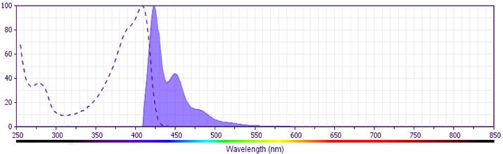
- For fluorochrome spectra and suitable instrument settings, please refer to our Multicolor Flow Cytometry web page at www.bdbiosciences.com/colors.
- BD Horizon Brilliant Violet 421 is covered by one or more of the following US patents: 8,158,444; 8,362,193; 8,575,303; 8,354,239.
- BD Horizon Brilliant Stain Buffer is covered by one or more of the following US patents: 8,110,673; 8,158,444; 8,575,303; 8,354,239.
- Please refer to http://regdocs.bd.com to access safety data sheets (SDS).
- Human donor specific background has been observed in relation to the presence of anti-polyethylene glycol (PEG) antibodies, developed as a result of certain vaccines containing PEG, including some COVID-19 vaccines. We recommend use of BD Horizon Brilliant™ Stain Buffer in your experiments to help mitigate potential background. For more information visit https://www.bdbiosciences.com/en-us/support/product-notices.
- For U.S. patents that may apply, see bd.com/patents.
- This reagent has been pre-diluted for use at the recommended Volume per Test. We typically use 1 × 10^6 cells in a 100-µl experimental sample (a test).
- An isotype control should be used at the same concentration as the antibody of interest.
Companion Products


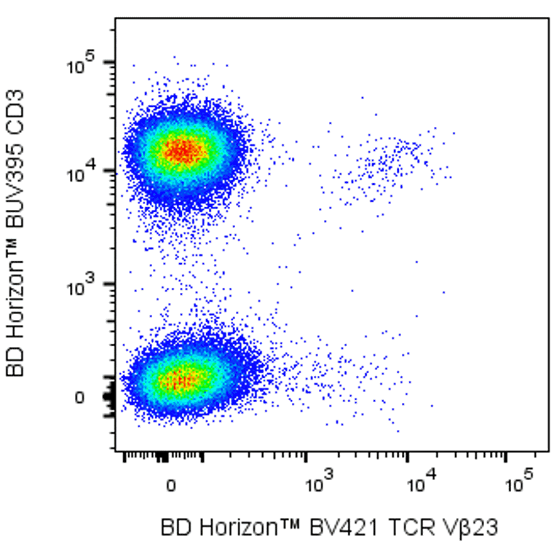
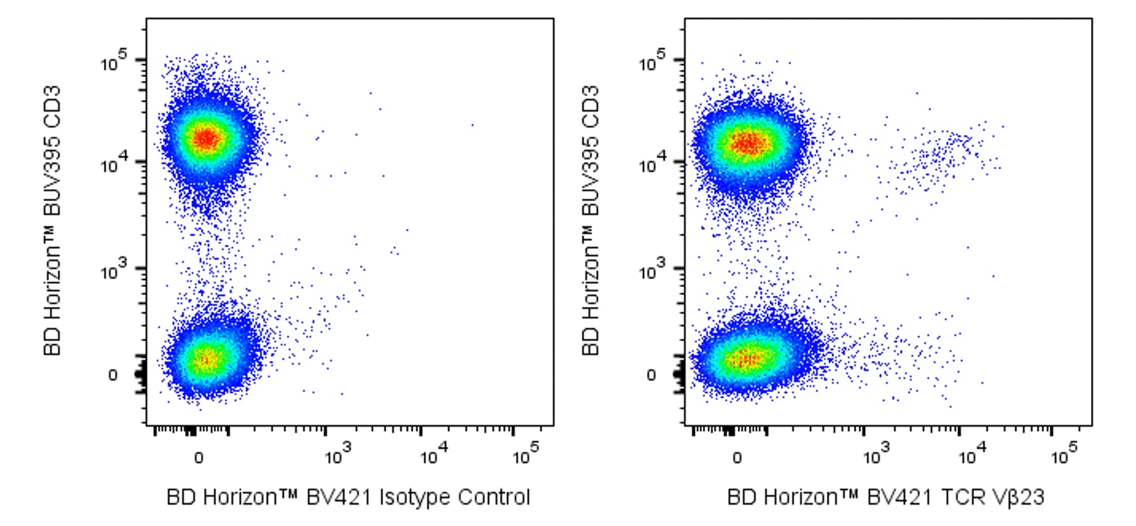
The AHUT7 monoclonal antibody specifically recognizes the human variable beta 23 region of the β subunit of the human αβ T cell receptor for antigen (TCR Vβ23). The TCR Vβ23 region is encoded by the TRBV13 gene segment. TCR Vβ23 is expressed on subsets of TCR αβ positive thymocytes and on ~0.3 to 5% of peripheral blood T cells including CD4+ and CD8+ T cells. The AHUT7 antibody is useful for multiparameter analyses designed to study the nature of TCR Vβ23-positive T cells including normal T cells as well as T cell clones, hybridomas, or tumor cell lines including H9 and HUT-78. It is also useful for analyzing TCR Vβ repertoires expressed by T cell populations collected from blood, tissues or other sources in health and disease models including inflammation, autoimmunity, responses to superantigens, tumors, and infectious diseases. The AHUT7 antibody can reportedly stimulate the proliferation of TCR Vβ23-positive T cells.
Development References (8)
-
Beres SB, Sylva GL, Barbian KD, et al. Genome sequence of a serotype M3 strain of group A Streptococcus: phage-encoded toxins, the high-virulence phenotype, and clone emergence.. Proc Natl Acad Sci U S A. 2002; 99(15):10078-83. (Biology). View Reference
-
Ghia P, Prato G, Stella S, Scielzo C, Geuna M, Caligaris-Cappio F. Age-dependent accumulation of monoclonal CD4+CD8+ double positive T lymphocytes in the peripheral blood of the elderly.. Br J Haematol. 2007; 139(5):780-90. (Clone-specific: Flow cytometry). View Reference
-
Giudice V, D'Addona M, Montuori N, Selleri C. The Value of Flow Cytometry Clonality in Large Granular Lymphocyte Leukemia.. Cancers (Basel). 2021; 13(18):4513. (Clone-specific: Flow cytometry). View Reference
-
Guilherme L, Faé KC, Oshiro SE, Tanaka AC, Pomerantzeff PM, Kalil J. T cell response in rheumatic fever: crossreactivity between streptococcal M protein peptides and heart tissue proteins.. Curr Protein Pept Sci. 2007; 8(1):39-44. (Biology). View Reference
-
Lavoie PM, Dumont AR, McGrath H, Kernaleguen AE, Sékaly RP. Delayed expansion of a restricted T cell repertoire by low-density TCR ligands.. Int Immunol. 2005; 17(7):931-41. (Clone-specific: Flow cytometry). View Reference
-
Pantaleo G, Soudeyns H, Demarest JF, et al. Evidence for rapid disappearance of initially expanded HIV-specific CD8+ T cell clones during primary HIV infection.. Proc Natl Acad Sci U S A. 1997; 94(18):9848-53. (Clone-specific: Fluorescence activated cell sorting). View Reference
-
Rebai N, Pantaleo G, Demarest JF, et al. Analysis of the T-cell receptor beta-chain variable-region (V beta) repertoire in monozygotic twins discordant for human immunodeficiency virus: evidence for perturbations of specific V beta segments in CD4+ T cells of the virus-positive twins.. Proc Natl Acad Sci U S A. 1994; 91(4):1529-33. (Clone-specific: Flow cytometry). View Reference
-
Romagné F, Kanagawa O, David-Ameline J, Peyrat MA, Bonneville M, Necker A. TCRBV23 specificity of two monoclonal antibodies revealed by a panel of human V beta chains expressed in mouse cells.. J Immunol Methods. 1995; 186(2):313-22. (Immunogen: Flow cytometry). View Reference
Please refer to Support Documents for Quality Certificates
Global - Refer to manufacturer's instructions for use and related User Manuals and Technical data sheets before using this products as described
Comparisons, where applicable, are made against older BD Technology, manual methods or are general performance claims. Comparisons are not made against non-BD technologies, unless otherwise noted.
For Research Use Only. Not for use in diagnostic or therapeutic procedures.