







Preparation And Storage


Recommended Assay Procedures
Cell Preparation and Staining Procedures for Fluorochrome-Conjugated Anti-Human FoxP3 Antibody
1. Bring the buffers to room temperature (RT) before use. Prepare working solutions of the BD Pharmingen Human FoxP3 Buffer Set
Cat. No. 560098 (For the buffer preparation, please see the Technical Data Sheet of Cat. No. 560098 buffer instructions for details).
2. Prepare human PBMC. Suspend the cells with BD Pharmingen™ Stain Buffer (FBS)* to ten million cells/ml.
3. Pipette appropriate amount of surface staining reagent to the bottom of each 12 × 75 mm tube.
4. Add 100 µl of cells per tube, vortex, incubate for 20 minutes at RT protected from light.
5. Add 2 ml of wash buffer. Centrifuge 250g for 10 minutes to pellet the cells and remove wash buffer.
6. To fix the cells, gently resuspend the cell pellet in residual volume of wash buffer and then add 2 ml of 1× Human FoxP3 Buffer A.
Vortex. Incubate for 10 minutes at RT in the dark.
7. Centrifuge 500g for 5 minutes, and remove fixative. Caution: Be aware the pellet is buoyant.
8. To wash cells, resuspend each cell pellet in 2 ml of BD Pharmingen Stain Buffer (FBS)*, and centrifuge 500g for 5 minutes. Remove
wash buffer.
9. To permeabilize the cells, gently resuspend pellet in residual volume of wash buffer and then add 0.5 ml of 1× working solution
Human FoxP3 Buffer C to each tube. Vortex. Incubate for 30 minutes at RT protected from light.
10. To wash cells, add 2 ml of BD Pharmingen™ Stain Buffer (FBS)* to each tube, centrifuge 500g for 5 minutes at RT. Remove buffer
and repeat wash step. Remove buffer.
11. Add conjugated FoxP3 antibody at appropriate concentrations to resuspend the pellet. Gently shake or vortex.
12. Incubate for 30 minutes in the dark at RT.
13. Repeat wash step #10.
14. Resuspend in wash buffer and analyze immediately.
Optional Add 300 µl of 1% formaldehyde in 1× PBS and store at 4°C. Analyze cells within 24 hours.
* We recommend using the BD Pharmingen™ Stain Buffer (FBS; Cat No. 554656) for all wash steps and covering tubes during incubation steps with caps or parafilm. We also recommend optimizing forward scatter and side scatter voltages to visualize lymphocytes as separate from debris, red cell ghosts and/or platelets before acquisition.
Product Notices
- This reagent has been pre-diluted for use at the recommended Volume per Test. We typically use 1 × 10^6 cells in a 100-µl experimental sample (a test).
- Source of all serum proteins is from USDA inspected abattoirs located in the United States.
- Please refer to www.bdbiosciences.com/us/s/resources for technical protocols.
- Cy is a trademark of Amersham Biosciences Limited. This conjugated product is sold under license to the following patents: US Patent Nos. 5,486,616; 5,569,587; 5,569,766; 5,627,027.
- Please observe the following precautions: Absorption of visible light can significantly alter the energy transfer occurring in any tandem fluorochrome conjugate; therefore, we recommend that special precautions be taken (such as wrapping vials, tubes, or racks in aluminum foil) to prevent exposure of conjugated reagents, including cells stained with those reagents, to room illumination.
- Caution: Sodium azide yields highly toxic hydrazoic acid under acidic conditions. Dilute azide compounds in running water before discarding to avoid accumulation of potentially explosive deposits in plumbing.
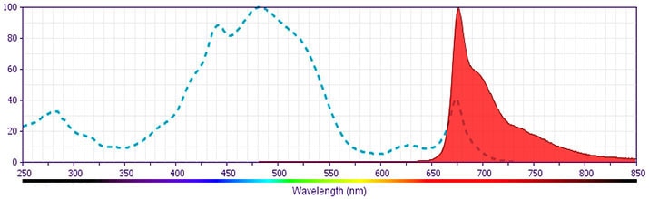
- PerCP-Cy5.5–labelled antibodies can be used with FITC- and R-PE–labelled reagents in single-laser flow cytometers with no significant spectral overlap of PerCP-Cy5.5, FITC, and R-PE fluorescence.
- PerCP-Cy5.5 is optimized for use with a single argon ion laser emitting 488-nm light. Because of the broad absorption spectrum of the tandem fluorochrome, extra care must be taken when using dual-laser cytometers, which may directly excite both PerCP and Cy5.5™. We recommend the use of cross-beam compensation during data acquisition or software compensation during data analysis.
- For fluorochrome spectra and suitable instrument settings, please refer to our Multicolor Flow Cytometry web page at www.bdbiosciences.com/colors.
- This product is subject to proprietary rights of Amersham Biosciences Corp. and Carnegie Mellon University and made and sold under license from Amersham Biosciences Corp. This product is licensed for sale only for research. It is not licensed for any other use. If you require a commercial license to use this product and do not have one return this material, unopened to BD Biosciences, 10975 Torreyana Rd, San Diego, CA 92121 and any money paid for the material will be refunded.
Companion Products

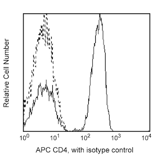
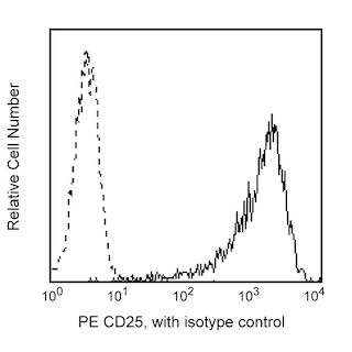
The 236A/E7 antibody reacts with the human FoxP3 (Forkhead box protein P3) transcription factor, a member of the forkhead or winged helix family of transcription factors. The expression of FoxP3, also known as Scurfin, IPEX and JM2, has been found to be associated with CD4+ CD25+ regulatory T cells and represents a specific marker for these cells. Flow cytometric analysis has shown that FoxP3 is expressed by the majority of CD4+ CD25+ (high) T cells in peripheral blood while less than half of CD4+ CD25+ (intermediate) cell population are FoxP3 positive. Approximately 5-10% of peripheral CD4+ cells are CD4+ CD25+ T regulatory cells. T regulatory cells are thought to play a critical role in the control of T cell mediated autoimmunity by suppressing the proliferation and cytokine production of other T cells. To support this hypothesis, it has been found that Foxp3 is mutated in scurfy (sf) mice. Cumulative evidence suggests that the 236A/E7 antibody recognizes epitopes from both isoforms of Exon 2 alternatively-spliced variants and is located in the region between Exon 2 and the Zn/LZ domains (aa 105-235).
Development References (8)
-
Alvaro T, Lejeune M, Salvado MT, et al. Outcome in Hodgkin's lymphoma can be predicted from the presence of accompanying cytotoxic and regulatory T cells. Clin Cancer Res. 2005; 11(4):1467-1473. (Clone-specific: Immunohistochemistry). View Reference
-
Brunkow ME, Jeffery EW, Hjerrild KA, et al. Disruption of a new forkhead/winged-helix protein, scurfin, results in the fatal lymphoproliferative disorder of the scurfy mouse. Nat Genet. 2001; 27(1):68-73. (Biology). View Reference
-
Fox BC, Bignone PA, Brown PJ, Banham AH. Defense of the clone: antibody 259D effectively labels human FOXP3 in a variety of applications. Blood. 2008; 111(7):3897-3899. (Clone-specific: Western blot). View Reference
-
Lennon G, Auffray C, Polymeropoulos M, Soares MB. Consortium: an integrated molecular analysis of genomes and their expression. Genomics. 1996; 33(1):151-152. (Biology). View Reference
-
Roncador G, Brown PJ, Maestre L, et al. Analysis of FOXP3 protein expression in human CD4+CD25+ regulatory T cells at the single-cell level. Eur J Immunol. 2005; 35(6):1681-1691. (Immunogen: Flow cytometry). View Reference
-
Roncador G, Garcia JF, Maestre L, et al. FOXP3, a selective marker for a subset of adult T-cell leukaemia/lymphoma. Leukemia. 2005; 19(12):2247-2253. (Clone-specific: Immunohistochemistry). View Reference
-
Wildin RS, Ramsdell F, Peake J, et al. X-linked neonatal diabetes mellitus, enteropathy and endocrinopathy syndrome is the human equivalent of mouse scurfy. Nat Genet. 2001; 27(1):18-20. (Biology). View Reference
-
Wolf D, Wolf AM, Rumpold H, et al. The expression of the regulatory T cell-specific forkhead box transcription factor FoxP3 is associated with poor prognosis in ovarian cancer. Clin Cancer Res. 2005; 11(23):8326-8331. (Biology). View Reference
Please refer to Support Documents for Quality Certificates
Global - Refer to manufacturer's instructions for use and related User Manuals and Technical data sheets before using this products as described
Comparisons, where applicable, are made against older BD Technology, manual methods or are general performance claims. Comparisons are not made against non-BD technologies, unless otherwise noted.
For Research Use Only. Not for use in diagnostic or therapeutic procedures.
Refer to manufacturer's instructions for use and related User Manuals and Technical Data Sheets before using this product as described.
Comparisons, where applicable, are made against older BD technology, manual methods or are general performance claims. Comparisons are not made against non-BD technologies, unless otherwise noted.
