





.png)


Preparation And Storage


Product Notices
- An isotype control should be used at the same concentration as the antibody of interest.
- Since applications vary, each investigator should titrate the reagent to obtain optimal results.
- Caution: Sodium azide yields highly toxic hydrazoic acid under acidic conditions. Dilute azide compounds in running water before discarding to avoid accumulation of potentially explosive deposits in plumbing.
- For fluorochrome spectra and suitable instrument settings, please refer to our Multicolor Flow Cytometry web page at www.bdbiosciences.com/colors.
- Please refer to www.bdbiosciences.com/us/s/resources for technical protocols.
Companion Products



.png?imwidth=320)
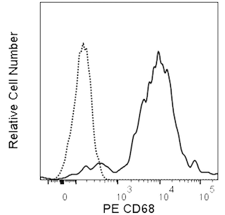
The FA/11 monoclonal antibody specifically binds to CD68 which is also known as Macrosialin, Lamp4 or Scard1. CD68 is a heavily glycosylated ~ 85-115 kDa type I transmembrane protein that belongs to the lysosomal-associated membrane protein (LAMP) family. Although it is expressed at the cell surface, it is predominantly expressed as an intracellular protein in late endosomes. CD68 is expressed by macrophages, Kupffer cells, histiocytes, microglia, and is considered a useful pan-macrophage marker. It is also expressed by dendritic cells, Langerhans cells and osteoclasts. CD68 expression is upregulated and its glycosylation patterns can be changed by proinflammatory agents. Although CD68 binds to oxidized low-density lipoproteins, its exact biological function is not well defined.

Development References (6)
-
Ashley JW, Shi Z, Zhao H, Li X, Kesterson RA, Feng X. Genetic ablation of CD68 results in mice with increased bone and dysfunctional osteoclasts.. PLoS ONE. 2011; 6(10):e25838. (Clone-specific: Flow cytometry, Immunofluorescence, Western blot). View Reference
-
Kurushima H, Ramprasad M, Kondratenko N, Foster DM, Quehenberger O, Steinberg D. Surface expression and rapid internalization of macrosialin (mouse CD68) on elicited mouse peritoneal macrophages.. J Leukoc Biol. 2000; 67(1):104-8. (Biology). View Reference
-
Rabinowitz SS, Gordon S. Macrosialin, a macrophage-restricted membrane sialoprotein differentially glycosylated in response to inflammatory stimuli.. J Exp Med. 1991; 174(4):827-36. (Clone-specific: Depletion, Immunocytochemistry, Immunohistochemistry, Immunoprecipitation, Radioimmunoassay, Western blot). View Reference
-
Ramprasad MP, Terpstra V, Kondratenko N, Quehenberger O, Steinberg D. Cell surface expression of mouse macrosialin and human CD68 and their role as macrophage receptors for oxidized low density lipoprotein.. Proc Natl Acad Sci USA. 1996; 93(25):14833-8. (Clone-specific: Flow cytometry). View Reference
-
Smith MJ, Koch GL. Differential expression of murine macrophage surface glycoprotein antigens in intracellular membranes.. J Cell Sci. 1987; 87 ( Pt 1):113-9. (Immunogen: Fluorescence microscopy, Immunofluorescence, Immunoprecipitation, Radioimmunoassay). View Reference
-
Song L, Lee C, Schindler C. Deletion of the murine scavenger receptor CD68.. J Lipid Res. 2011; 52(8):1542-50. (Clone-specific: Flow cytometry). View Reference
Please refer to Support Documents for Quality Certificates
Global - Refer to manufacturer's instructions for use and related User Manuals and Technical data sheets before using this products as described
Comparisons, where applicable, are made against older BD Technology, manual methods or are general performance claims. Comparisons are not made against non-BD technologies, unless otherwise noted.
For Research Use Only. Not for use in diagnostic or therapeutic procedures.
Refer to manufacturer's instructions for use and related User Manuals and Technical Data Sheets before using this product as described.
Comparisons, where applicable, are made against older BD technology, manual methods or are general performance claims. Comparisons are not made against non-BD technologies, unless otherwise noted.
