Old Browser
Looks like you're visiting us from {countryName}.
Would you like to stay on the current country site or be switched to your country?


.png)

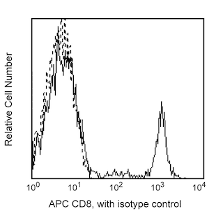
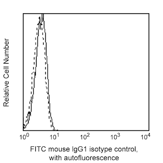
Expression of granzyme B by CD8 positive and CD8 negative peripheral blood mononuclear cells. Whole human blood was lysed with PharmLyse™ Lysing buffer (Cat. No. 555899) prior to staining with GB11. The lysed human blood was subsequently fixed, permeablilized and stained with APC- conjugated mouse anti-human CD8 (APC- RPA-T8, Cat. No. 555369) and either mouse anti-human granzyme B antibody (FITC- GB11, Cat. No. 558132), (filled histograms) or immunoglobulin isotype control (FITC- MOPC-21, Cat. No. 555909), (empty histograms) by using Pharmingen's staining protocol. To demonstrate specificity of staining, the binding of FITC-GB11 was blocked by preincubation of the fixed/permeabilized cells with an excess of unlabelled GB11 antibody (5 µg, data not shown) prior to staining. The histograms in the figure were derived from CD8-positive (Left panel) or CD8 -negative (Right panel) gated events.
.png)

BD Pharmingen™ FITC Mouse anti-Human Granzyme B
.png)
Regulatory Status Legend
Any use of products other than the permitted use without the express written authorization of Becton, Dickinson and Company is strictly prohibited.
Preparation And Storage
Product Notices
- This reagent has been pre-diluted for use at the recommended Volume per Test. We typically use 1 × 10^6 cells in a 100-µl experimental sample (a test).
- Please refer to www.bdbiosciences.com/us/s/resources for technical protocols.
- For fluorochrome spectra and suitable instrument settings, please refer to our Multicolor Flow Cytometry web page at www.bdbiosciences.com/colors.
- Caution: Sodium azide yields highly toxic hydrazoic acid under acidic conditions. Dilute azide compounds in running water before discarding to avoid accumulation of potentially explosive deposits in plumbing.
- Source of all serum proteins is from USDA inspected abattoirs located in the United States.
Companion Products


The GB11 antibody specifically reacts with human granzyme B, a serine protease of approximately 32 kDa. Granzyme B is stored in the granules of cytotoxic T lymphocytes and NK cells along with the pore-forming protein perforin. In the classic model of target cell lysis, perforins create holes in the target cell membrane allowing entrance of granzymes. Granzyme B has been shown to act on specific substrates including caspase-3, -7, -9, and -10 which in turn give rise to enzymes that mediate apoptosis. Granzyme B may also be involved in the hydrolysis of extracellular matrix components. Detectable levels of granzyme B have been detected in sera from healthy volunteers. The immunogen used to generate the GB11 hybridoma was human granzyme B isolated from an NK cell line.

Development References (9)
-
Hamann D, Baars PA, Rep MH. Phenotypic and functional separation of memory and effector human CD8+ T cells. J Exp Med. 1997; 186(9):1407-1418. (Clone-specific: Flow cytometry). View Reference
-
Poe M, Blake JT, Boulton DA. Human cytotoxic lymphocyte granzyme B. Its purification from granules and the characterization of substrate and inhibitor specificity. J Biol Chem. 1991; 266(1):98-103. (Biology). View Reference
-
Ronday HK, van der Laan WH, Tak PP et al. Human granzyme B mediates cartilage proteoglycan degradation and is expressed at the invasive front of the synovium in rheumatoid arthritis. Rheumatology (Oxford). 2001; 40:55-61. (Biology). View Reference
-
Smyth MJ, Kelly JM, Sutton VR et al. Unlocking the secrets of cytotoxic granule proteins. J Leukoc Biol. 2001; 70:18-29. (Biology). View Reference
-
Spaeny-Dekking EH, Hanna WL, Wolbink AM et al. Extracellular granzymes A and B in humans: detection of native species during CTL responses in vitro and in vivo. J Immunol. 1998; 160:3610. (Biology). View Reference
-
Trapani JA, Klein JL, White PC, and Dupont B. Molecular cloning of an inducible serine esterase gene from human cytotoxic lymphocytes. Proc Natl Acad Sci U S A. 1988; 5:6924-6928. (Biology). View Reference
-
Trapani JA, Smyth MJ, Apostolidis VA, Dawson M, and Browne KA. Granule serine proteases are normal nuclear constituents of natural killer cells. J Biol Chem. 1994; 269:18359-18365. (Biology). View Reference
-
Wever PC, Van Der Vliet HJ, Spaeny LH . The CD8+ granzyme B+ T-cell subset in peripheral blood from healthy individuals contains activated and apoptosis-prone cells. Immunology. 1998; 93(3):383-389. (Clone-specific: Flow cytometry). View Reference
-
ten Berge IJ, Wever PC, Rentenaar RJ. Selective expansion of a peripheral blood CD8+ memory T cell subset expressing both granzyme B and L-selectin during primary viral infection in renal allograft recipients. Transplant Proc. 1998; 30(8):3975-3977. (Biology). View Reference
Please refer to Support Documents for Quality Certificates
Global - Refer to manufacturer's instructions for use and related User Manuals and Technical data sheets before using this products as described
Comparisons, where applicable, are made against older BD Technology, manual methods or are general performance claims. Comparisons are not made against non-BD technologies, unless otherwise noted.
For Research Use Only. Not for use in diagnostic or therapeutic procedures.
Refer to manufacturer's instructions for use and related User Manuals and Technical Data Sheets before using this product as described.
Comparisons, where applicable, are made against older BD technology, manual methods or are general performance claims. Comparisons are not made against non-BD technologies, unless otherwise noted.
Report a Site Issue
This form is intended to help us improve our website experience. For other support, please visit our Contact Us page.